寄生蠕虫類の分類体系.第Ⅲ巻.脊椎動物の線虫類 原図 8/10
きせいぜんちゅうるいのぶんるいたいけい.だいさんかん.せきついどうぶつのせんちゅうるい げんず じゅうぶんのはち
概要
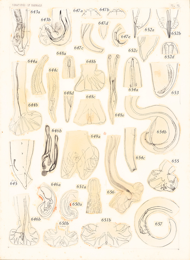
Plate 71.
Fig. 643. Pseudostenurus sunamevi Yamaguti, 1951. a. Posterior extremity of female. b. Posterior extremity of male.
Fig. 644. Heligmosomum costellatum (Duj., 1845). After Travassos and Darriba, 1929. a. Anterior extremity, lateral view. b. Posterior extremity of male. c. Posterior extremity of female.
Fig. 645. Paradujardinia halicoris (Owen, 1833); posterior extremity of male. After Yamaguti, 1941.
Fig. 646. Tricholinstowia linstowi (Travassos, 1918). a. Posterior extremity of female. b. Posterior extremity of male.
Fig. 647. Setaria equina (Abildg., 1789). After Yorke and Maplestone, 1926. a. Head, lateral view. b. Head, ventral view. c. Posterior extremity of female. d. Tail of female, dorsal view. e. Posterior extremity of male.
Fig. 648. Filarinema flagrifer Mönnig, 1929. a. Head. b—c. Bursa. d. Posterior extremity of female. e. Spicules.
Fig. 649. Spiculopteragia spiculoptera (Guschanskaja, 1931). a. Bursa. b. Spicules.
Fig. 650. Longistriata noviberiae Dikmans, 1935. a. Posterior extremity of female. b. Bursa.
Fig. 651. Brevistriata skrjabini (Schulz et Lubimow, 1932). a. Anterior extremity. b. Bursa.
Fig. 652. Filaroides mustelarum (van Beneden, 1858) = Filaroides martis (Werner 1782). After Cameron, 1927. a—b. Posterior extremity of male. c. Posterior extremity of female. d. Head.
Fig. 653. Filaroides martis (Werner, 1782); posterior extremity of male. After Petrow, 1928.
Fig. 654. Suifilaria suis Ortlepp, 1937. a. Anterior extremity. b. Posterior extremity of male. c. Posterior extremity of female.
Fig. 655. Heligmoskrjabinia skrjabini Freitas et Lent, 1937; bursa.
Fig. 656. Stilestrongylus stilesi Freitas, Lent et Almeida, 1937; posterior extremity of male.
Fig. 657. Molinema diacantha (Molin, 1858); posterior extremity of male.
After Freitas & Lent, 1939.
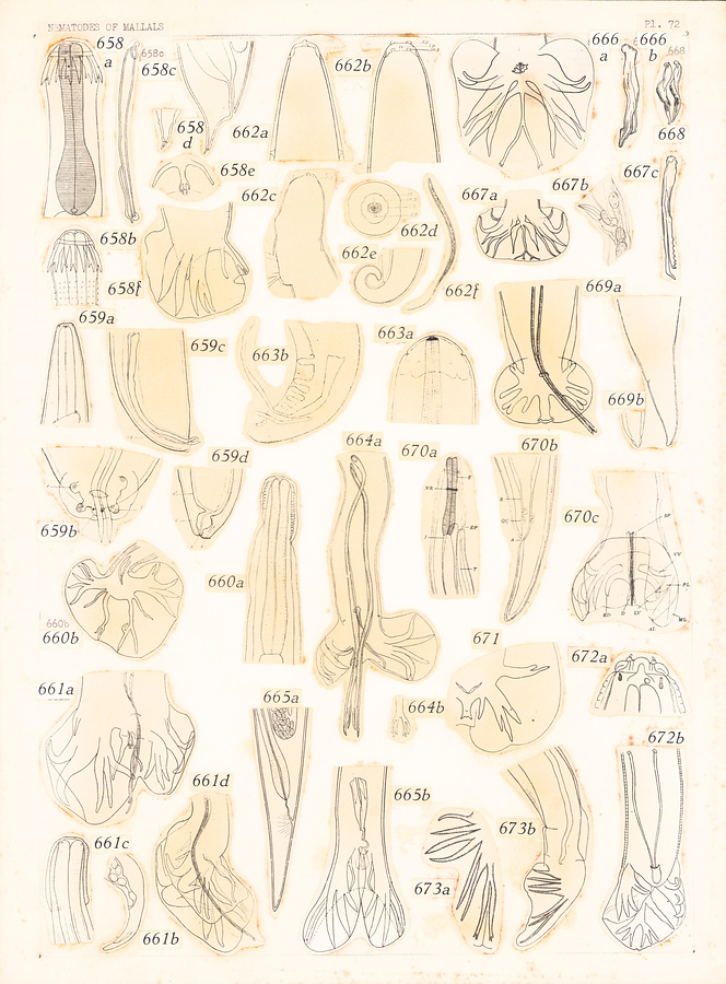
Plate 72.
Fig. 658. Spinostrongylus spinosus (Boulenger, 1926). After Boulenger in Travassos, 1937. a—b. Anterior extremity c. Spicule and gubernaculum. d. Posterior extremity of female. e. Dorsal ray. f. Bursa.
Fig. 659. Halocercus delphini Baylis et Daubney, 1925. a. Head. b—c. Posterior extremity of male. d. Posterior extremity of female.
Fig. 660. Pseudoheligmosomum howelli (Vigueras, 1934). After Vigueras in Travassos, 1937. a. Anterior extremity. b. Posterior extremity of male.
Fig. 661. Nematospiroides dubia Baylis, 1926. After Schulz in Travassos, 1937. a—b. Posterior extremity of male. c. Head. d. Posterior extremity of female.
Fig. 662. Dracunculus medinensis (Linn., 1758). After Yorke and Maplestone, 1926. a. Anterior extremity of female, ventral view. b. Anterior extremity of young female, lateral view. c. Anterior extremity of gravid female. d. Head, end-on view. e. Posterior extremity of female. f. Larva.
Fig. 663. Tawila tawila Khalil, 1932. a. Head. b. Posterior extremity of male.
Fig. 664. Trifurcata major Travassos, 1937. a. Posterior extremity of male. b. Dorsal ray.
Fig. 665. Leiperiatus hopkeni (Leiper, 1910). After Travassos, 1937. a. Posterior extremity of female. b. Posterior extremity of male.
Fig. 666. Cooperioides kenyensis Daubney, 1933. a. Bursa. b. Spicule.
Fig. 667. Paracooperia serrata (Mönnig, 1931). After Mönnig in Travassos, 1937. a. Bursa. b. Vulvar region. c. Spicule.
Fig. 668. Cooperia Pectinata Ransom, 1907; Spicules.
Fig. 669. Bronchostrongylus subcrenatus (Raill. et Henry, 1913). After Raill. & Henry, 1913. a. Posterior extremity of male. b. Posterior extremity of female.
Fig. 670. Moguranema nipponicum Yamaguti, 1941. a. Anterior extremity, lateral view. b. Posterior extremity of female. c. Posterior extremity of male.
Fig. 671. Rodentocaulus ondatrae Schulz, Orloff et Kutass, 1933; bursa.
Fig. 672. Paraquilonia brumpti Neveu-Lemaire, 1924. After Leveu-Lemaire in Yorke and Maplestone, 1926. a. Head, lateral view. b. Posterior extremity of male.
Fig. 673. Mazamanema longibursatum Cameron, 1935. a. Posterior extremity of male. b. Bursal rays.
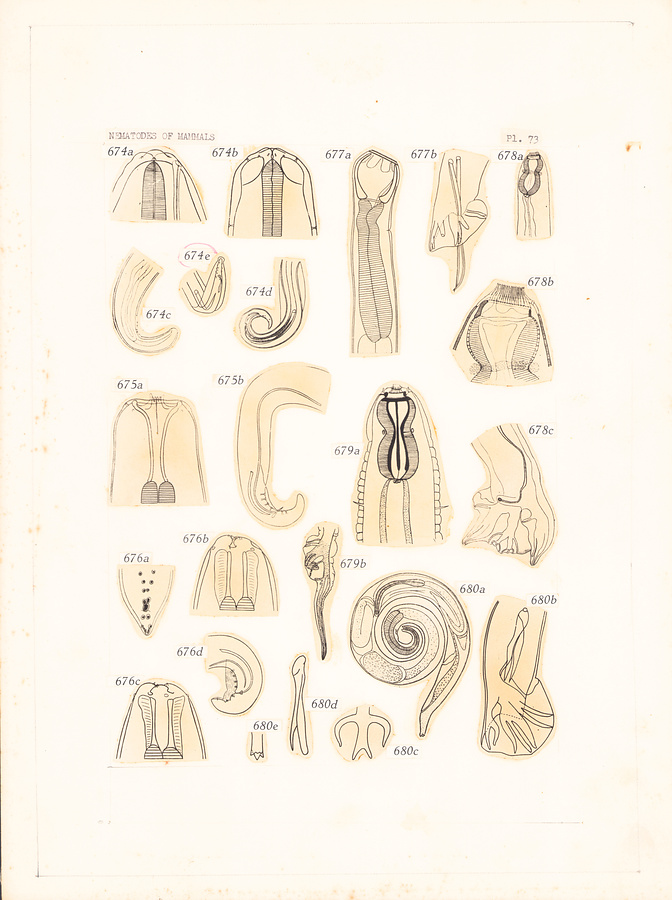
Plate 73.
Fig. 674. Filaria martis Gmelin, 1790. After Yorke and Maplestone, 1926. a. Head, lateral view. b. Head, ventral view. c. Posterior extremity of female. d—e. Posterior extremity of male.
Fig. 675. Cylicospirura subaequalis (Molin, 1860). After Yorke and Maplestone, 1926. a. Anterior extremity, ventral view. b. Posterior extremity of male.
Fig. 676. a. Posterior extremity of male of Protospirura numidica Seurat, 1914. After Seurat, 1914. b. Head of Protospirura ascaroidea Hall, 1916, ventral view. c. Head of same, lateral view. c. Posterior extremity of male of Protospirura ascaroidea Hall, 1916. After Y. & M., 1926.
Fig. 677. Chabertia ovina (Fabricius, 1794). After Yorke and Maplestone, 1926. a. Anterior extremity, lateral view. b. Posterior extremity of male.
Fig. 678. Khalilia rhinocerotis Neveu-Lemaire, 1924. a. Anterior extremity, lateral view. b. Head, lateral view. c. Posterior extremity of male.
Fig. 679. Amiroides pileata (Raill., Henry et Bauche, 1914). After Khalil in Yorke and Maplestone, 1926. a. Anterior extremity, ventral view. b. Bursa.
Fig. 680. Ollulanus tricuspis Leuckart, 1865. After Cameron in Travassos, 1937. a. Female. b. Posterior extremity of male. c. Dorsal ray. d. Spicule. e. Tail end of female.
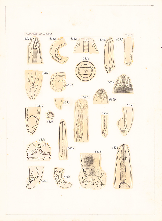
Plate 74.
Fig. 681. Streptopharagus armatus Blanc, 1912. After Yorke and Maplestone, 1926. a. Posterior extremity of male. b. Anterior extremity, ventral view. c. Posterior extremity of male.
Fig. 682. Lagochilascaris minor Leiper, 1909. After Leiper in Yorke and Maplestone, 1926. a. Posterior extremity of male. b. Egg. c. Head, ventral view.
Fig. 683. Dipetalonema gracile (Rud., 1809). After Yorke and Maplestone, 1926. a. Head, ventral view. b. Anterior extremity of female. c. Head, end-on view. d. Posterior extremity of male. e. Tail end of female.
Fig. 684. Trachypharynx nigeriae Leiper, 1911; anterior extremity, lateral view. After Leiper in Yorke and Maplestone, 1926.
Fig. 685. Parafilaria multipapiIlosa (Condamine et Drouilly, 1878). After Railliet in Yorke and Maplestone 1926.
a—b. Head of female, ventral and lateral view, respectively. c—d. Posterior extremity of male. e. Posterior extremity of female.
Fig. 686. Loa loa (Guyot, 1778). After Yorke and Maplestone, 1926. a. Anterior extremity, lateral view. b. Posterior extremity of male. c. Posterior extremity of female.
Fig. 687. Rattostrongylus cantonensis (Chen, 1935). a. Anterior extremity. b. Bursa.
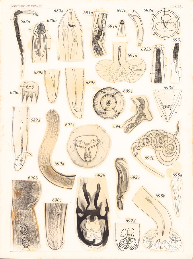
Plate 75.
Fig. 688. Acanthoxyurus anomaluri Sandground, 1928. a. Female. b. Anterior extremity, dorsolateral view. c. Head, end-on view.
Fig. 689. Pseudofilaria pertenuis (Rodhain, 1919). After Sandground, 1935. a. Anterior extremity of female, lateral view. b. Posterior extremity of female. c—d. Posterior extremity of male. e. Head, end-on view.
Fig. 690. Anatrichosoma cynamolgi Smith et Chitwood, 1954. a. Anterior extremity of female. b. Vulvar region. c. Posterior extremity of male.
Fig. 691. Anoplostrongylus paradoxus (Travassos, 1918). After Travassos, 1921. a. Vulvar region. b. Head. c. Posterior extremity of female. d. Posterior extremity of male.
Fig. 692. Böhmiella perichitinea Gebauer, 1932. a. Head, end-on view. b. Posterior extremity of male. c. Spicule. d. Bursa.
Fig. 693. Terranova azarasi (Yamaguti et Arima, 1942). a. Head end-on view. b. Anterior extremity, dorsal view. c. Ventricular region. d. Posterior extremity of male.
Fig. 694. Nematodirella longispiculata Yorke et Maplestone, 1926. After Skrjabin in Travassos, 1937. a. Vulvar region. b. Male.
Fig. 695. Nochtia nochti Travassos et Vogelsang, 1929. a. Posterior extremity of male. b. Proximal end of spicules.
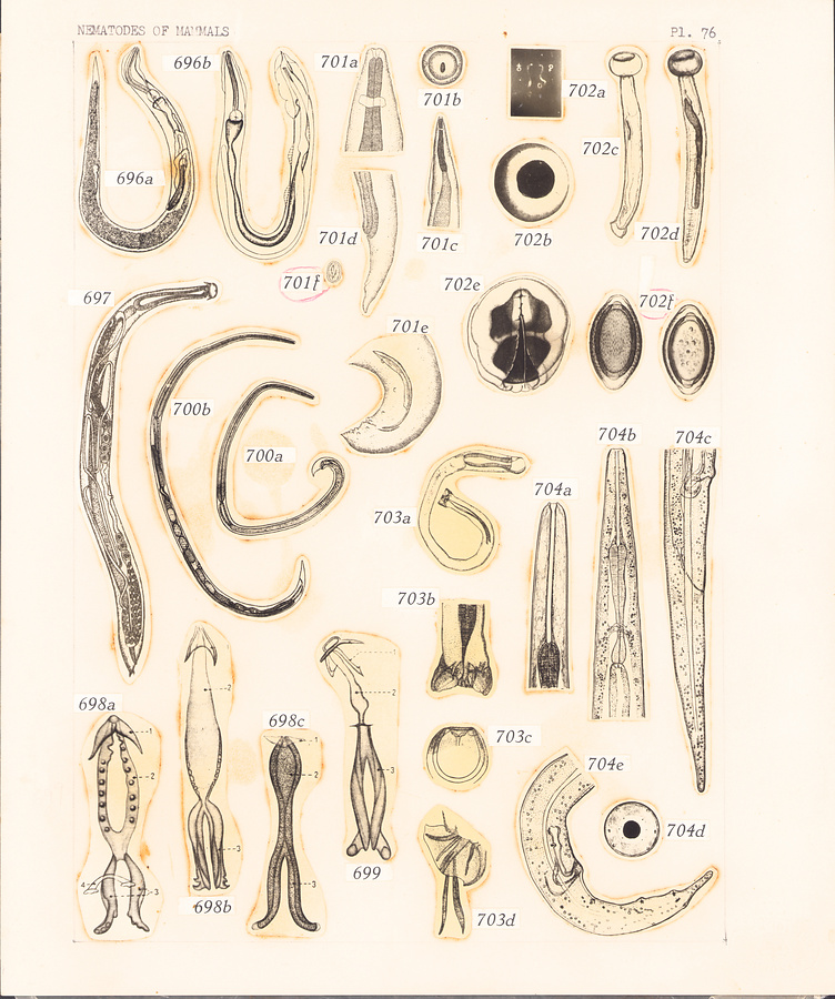
Plate 76.
Fig. 696. Dermatopallarya baylisi Skrjabin, 1924. After Skrjabin in Yorke and Maplestone, 1926. a. Female. b. Male.
Fig. 697. Histiostrongylus coronatus Molin, 1861; female. After Molin in Yorke and Maplestone, 1926.
Fig. 698. Gubernacula of Protostrongylus spp. 1. Captum, 2. Corpus, 3. Crura. a. Protostrongylus kochi (Schulz, Orlow et Kutass, 1933) = P. rufescens. b. Protostrongylus raillieti Schulz, Orlow et Kutass, 1933. c. Protostrongylus leuckarti Schulz, Orlow et Kutass, 1933.
Fig. 699. Gubernaculum of Cystocaulus nigrescens (Jerke, 1911). After Schulz, Orlow and Kutass, 1933.
Fig. 700. Parastrongyloides winchesi Morgan, 1928. a. Male. b. Female.
Fig. 701. Metathelazia californica Skinker, 1931. a. Head. b. Head, end-on view. c. Anterior extremity, lateral view. d. Posterior extremity of female. e. Posterior extremity of male. f. Egg.
Fig. 702. Soboliphyme baturini Petrow, 1930. a. Male and female. b. Head, end-on view. c. Male. d. Female. e. Bursa, end-on view. f. Egg, surficial view and optical section, respectively
Fig. 703. Ransomus rodentorum Hall, 1916. After Hall in Yorke and Maplestone, 1926. a. Male. b. Posterior extremity of male. c. Head, dorsal view. d. Bursa.
Fig. 704. Longibucca lasiura McIntosh et Chitwood, 1934. a—b. Anterior extremity. c. Posterior extremity of female. d. Head, end-on view. e. Posterior extremity of male.
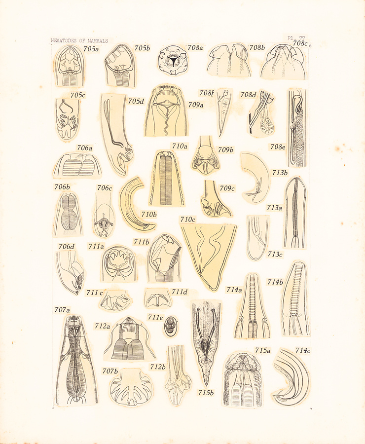
Plate 77.
Fig. 705. Gaigeria pachyscelis Railliet et Henry, 1910. After Yorke and Maplestone, 1926. a. Head, dorsal view. b. Head, lateral view. c—d. Posterior extremity of male.
Fig. 706. Hoplodontophorus flagellum Ehrenberg in Hemprich and Ehrenberg, 1928. After Turner in Yorke and Maplestone, 1926. a. Head, lateral view. b. Anterior extremity, ventral view. c—d. Posterior extremity of male.
Fig. 707. Ternidens deminutus (Railliet et Henry, 1905). After R. & H. in Yorke and Maplestone, 1926. a. Anterior extremity, ventral view. b. Bursa.
Fig. 708. Pseudocruzia orientalis (Maplestone, 1930). a. Head, end-on view. b. Head, lateral view. c. Head, dorsal view. d. Anal region. e. Vulvar region. f. Tail of female.
Fig. 709. Poteriostomum imparidentatum Quiel, 1919. After Yorke and Macfie in Yorke and Maplestone, 1926. a. Head, ventral view. b=c. Bursa.
Fig. 710. Thelazia rhodesii (Desmarest, 1828). After Yorke and Maplestone, 1926. a. Anterior extremity, ventral view. b. Posterior extremity of male. c. Posterior extremity of female.
Fig. 711. Ancylostoma duodenale (Dubini, 1843.) a. Head, dorsal view. b. Head, lateral view. c—d. Bursa. e. Egg.
Fig. 712. Buissonia rhinocerotis Neveu-Lemaire, 1924. After Neveu-Lemaire in Yorke and Maplestone, 1926. a. Head, lateral view. b. Posterior extremity of male.
Fig. 713. Dirofilaria immitis (Leidy, 1856). After Yorke and Maplestone, 1926. a. Anterior extremity, ventral view. b. Posterior extremity of male. c. Posterior extremity of female.
Fig. 714. Physocephalus sexalatus (Molin, 1860); After Yorke and Maplestone, 1926. a. Anterior extremity, ventral view. b. Anterior extremity, lateral view. c. Posterior extremity of male.
Fig. 715. Paraspidodera uncinata (Rud., 1819). After Travassos in Yorke and Maplestone, 1926. a. Anterior extremity, dorsal view. b. Posterior extremity of male.
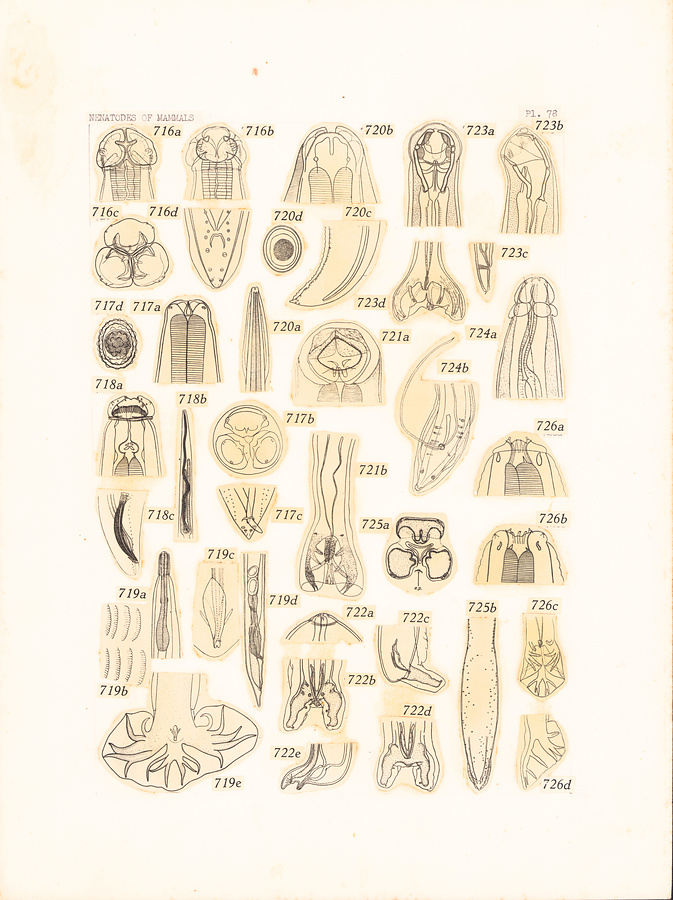
Plate78.
Fig. 716. Parascaris equorum (Goeze, 1782). After Yorke and Maplestone, 1926. a. Head, ventral view. b. Head, dorsal view. c. Head, end-on view. d. Posterior extremity of male.
Fig. 717. Ascaris lumbricoides Linné, 1758. After Yorke and Maplestone, 1926. a. Anterior extremity, ventral view. b. Head, end-on view. c. Posterior extremity of male. d. Egg.
Fig. 718. Crossophorus collaris Hemprich et Ehrenb., 1828. After Yorke and Maplestone, 1926. a. Head, dorsal view. b. Anterior extremity. c. Posterior extremity of male.
Fig. 719. Heligmodendrium hassalli (Price, 1929). a. Anterior extremity. b. Striated cuticle. c. Spicules and gubernaculum. d. Posterior extremity of female. e. Posterior extremity of male.
Fig. 720. Toxascaris leonina (Linstow, 1902). After Yorke and Maplestone, 1926. a. Anterior extremity, ventral view. b. Head, dorsal view. c. Posterior extremity of male. d. Egg.
Fig. 721. Brachyclonus indicus Railliet et Henry, 1910. After Khalil in Yorke and Maplestone, 1926. a. Head, dorsal view. b. Posterior extremity of male.
Fig. 722. Pseudalius inflexus (Rud., 1809). After Baylis and Daubney in Yorke and Maplestone, 1926. a. Head, lateral view. b—d. Posterior extremity of male. e. Posterior extremity of female.
Fig. 723. Arthrocephalus gambiensis Ortlepp, 1925. After Ortlepp in Yorke and Maplestone, 1926. a. Anterior extremity, dorsal view. b. Anterior extremity, lateral view. c. Posterior extremity of female. d. Posterior extremity of male.
Fig. 724. Squamanema bonnei Thiel, 1925. After Thiel in Yorke and Maplestone, 1926. a. Anterior extremity. b. Posterior extremity of male.
Fig. 725. Filocapsularia rosmari (Baylis, 1916). After Baylis in Yorke and Maplestone, 1926. a. Head, end-on view. b. Posterior extremity of male.
Fig. 726. Quilonia africana Lane, 1921. After Yorke and Maplestone, 1926. a. Head, lateral view. b. Head, ventral view. c—d. Bursa.
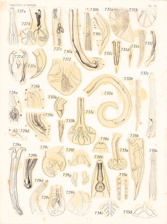
Plate 79.
Fig. 727. Travassostrongylus callis (Travassos, 1914). After Orloff, 1933. a. Anterior extremity. b. Posterior extremity of male. c. Female terminalia. d. Spicules. e. Posterior extremity of female. f. Bursa.
Fig. 728. Oxyuronema atelophorum Kreis, 1932. a. Posterior part of esophagus. b. Head. c. Posterior extremity of male. d. Tail spike of male.
Fig. 729. Avellaria avellari Freitas et Lent, 1934. a. Spicules. b. Distal portion of spicules. c. Bursa. d. Anterior extremity. e. Posterior extremity of female. f—h. Bursal rays.
Fig. 730. Neometastrongylus buechii Kreis, 1944. a. Posterior extremity of male. b. Anterior extremity, ventral view. c. Posterior extremity of female.
Fig. 731. Asymmetricostrongylus asymmetricus (Cameron, 1926). After Nagaty, 1938. a—b. Bursa. c. Spicules. d. Gubernaculum. e. Posterior extremity of female. f. Spicule and gubernaculum, lateral view
Fig. 732. Ackertia burgosi (De la Barrera, 1926). After Freitas, Lent & Almeida, 1937. a. Posterior extremity of male. b. Posterior extremity of f male. c. Anterior extremity of female, lateral view.
Fig. 733. Heligmostrongylus sedecimradiatus (Linstow, 1899). After Travassos in Yorke and Maplestone, 1926. a. Posterior extremity of old female. b. Posterior extremity of young female. c. Posterior extremity of male.
Fig. 734. Eucyathostomum longesubulatum Molin, 1861. After Cameron, 1936 a. Head, lateral view. b. Anterior extremity, lateral view. c. Dorsal ray.
Fig. 735. Trichotravassosia travassosi Lent et Freitas, 1938. a. Posterior extremity of male. b. Posterior extremity of female. c. Anterior extremity. d. Dorsal ray. e. Scale-like cuticle.
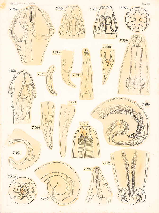
Plate 80.
Fig. 736. Stammerinema soricis (Tiner, 1951). After Osche, 1955. a—b. Anterior extremity of female. c. Shorter spicule. d—e. Posterior extremity of male. f. Posterior extremity of female.
Fig. 737. Labiobulura peramelis Baylis, 1930. a. Head, end-on view. b. Posterior extremity of male. c. Head, lateral view.
Fig. 738. Sexansodera biansata (Railliet et Henry, 1913). After Proenqa, 1937, in Skrjabin et al., 1951. a. Head, ventral view. b. Head, lateral view. c—d. Posterior extremity of male. e. Anterior extremity, ventral view.
Fig. 739. Skyjabinocercina petrowi Machulskii, 1952. a. Head, end-on view. b. Anterior extremity, lateral view. c. Posterior extremity of male.
Fig. 740. Skrjabingylus nasicola (Leuckart, 1842). After Petrow, 1927. a. Anterior extremity, lateral view. b. Posterior extremity of male.
所蔵館のウェブサイトで見る
公益財団法人 目黒寄生虫館文化庁 〒602-8959 京都府京都市上京区下長者町通新町西入藪之内町85番4
(C) The Agency for Cultural Affairs