日本の寄生蠕虫相の研究,第33部.魚類の線虫類 II.原図
にほんのきせいぜんちゅうそうのけんきゅう,だいさんじゅうさんぶ.ぎょるいのせんちゅうるいに.げんず

概要
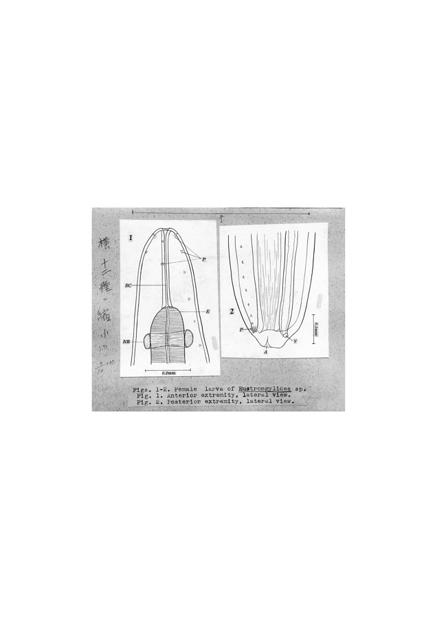
Figs. 1-2. Female larva of Eustrongylides sp.: Fig. 1. Anterior extremity, lateral view., Fig. 2. Posterior extremity, lateral view.
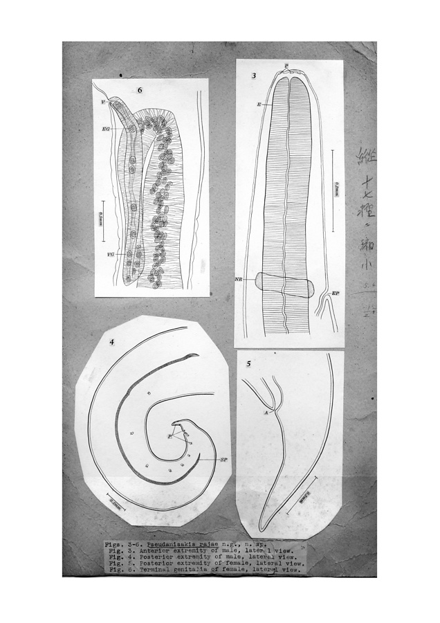
Figs. 3-6. Pseudanisakis rajae n. g., n. sp.: Fig. 3. Anterior extremity of male, lateral view., Fig. 4. Posterior extremity of male, lateral view.
Fig. 5. Posterior extremity of female, lateral view.
Fig. 6. Terminal genitalia of female, lateral view.
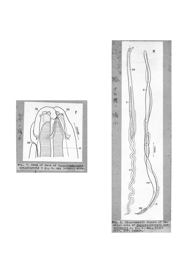
Fig. 7. Head of male of Paranisakiopsis coelorhynchi n. g., n. sp. lateral view.
Fig. 8. Diagrammatic figure of genital sets of Paranisakiopsis coelorhynchi n. g., n. sp., right male, left female.
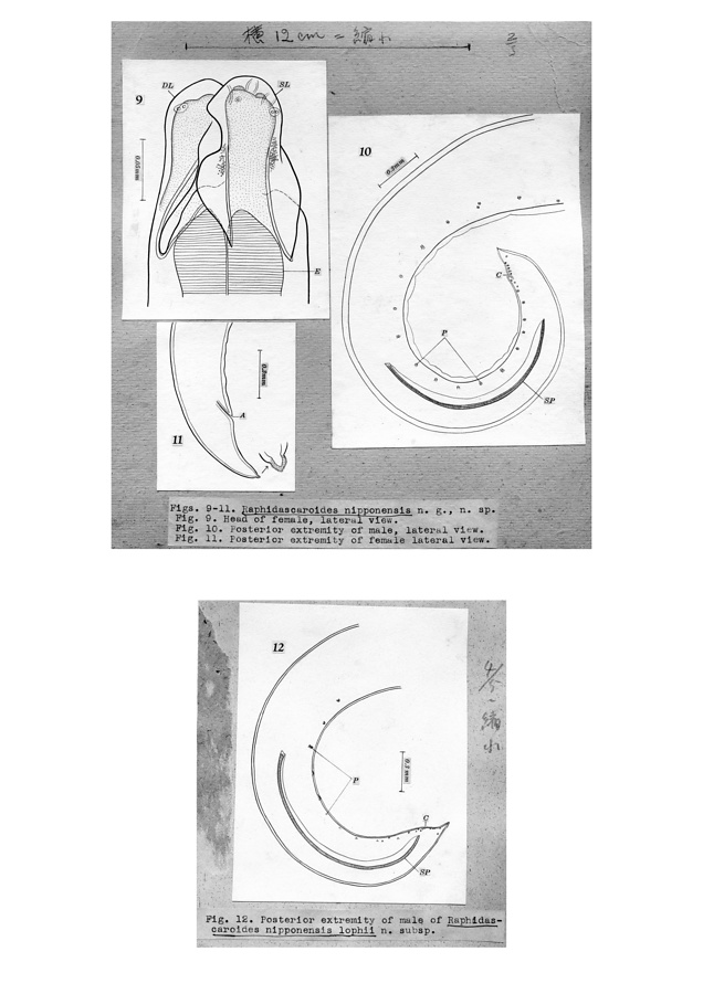
Figs. 9-11. Raphidascaroides nipponensis n. g., n. sp.: Fig. 9. Head of female, lateral view., Fig. 10. Posterior extremity of male, lateral view., Fig. 11. Posterior extremity of female, lateral view.
Fig. 12. Posterior extremity of male of Raphidascaroides nipponensis lophii n. subsp.
Fig. 13. Head of male of Porrocaecum cephaloscylii n. sp., ventral view.
Fig. 14. Head of female of Contracaecum epinepheli n. sp., lateral view.
Fig. 15. Head of female of Contracaecum seriolae n. sp., lateral view.
Figs. 16-17. Female of Contracaecum paralichthydis n. sp.: Fig. 16. Head, lateral view., Fig. 17. Tail, lateral view.
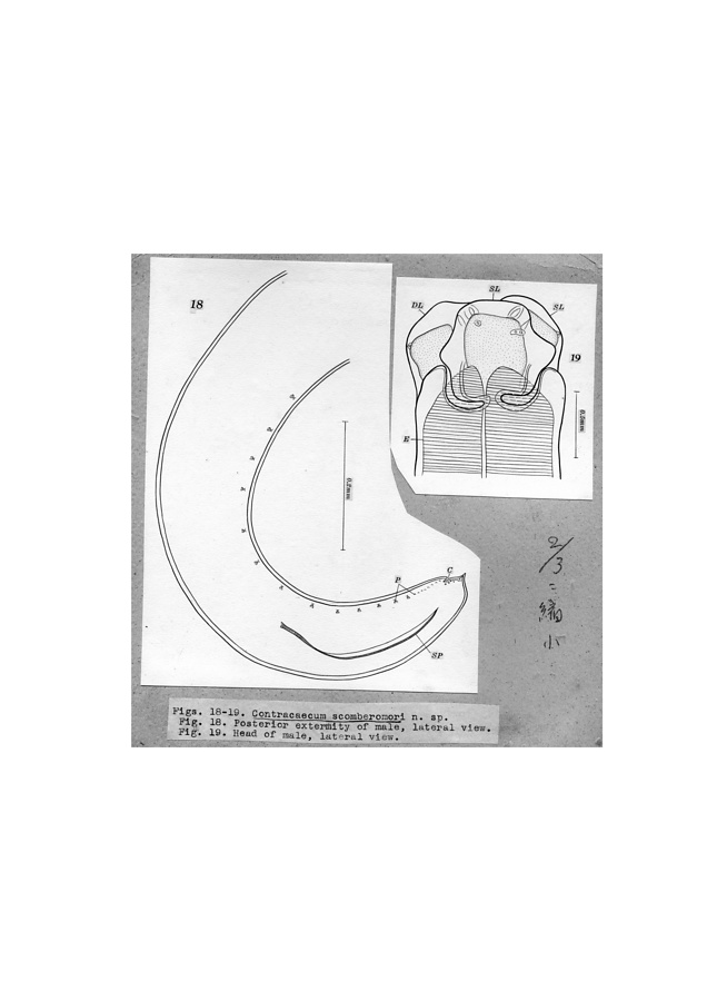
Figs. 18-19. Contracaecum scomberomori n. sp.: Fig. 18. Posterior extremity of male, lateral view., Fig. 19. Head of male, lateral view.
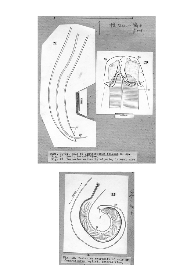
Figs. 20-21. Male of Contracaecum coiliae n. sp.: Fig. 20. Head, lateral view., Fig. 21. Posterior extremity of male, lateral view.
Fig. 22. Posterior extremity of male of Contracaecum baylisi, lateral view.
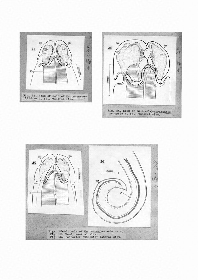
Fig. 23. Head of male of Contracaecum ilishae n sp., ventral view.
Fig. 24. Head of Contracaecum baylisi n. sp., ventral view.
Figs. 25-26. Male of Contracaecum saba n. sp.: Fig. 25. Head, ventral view., Fig. 26. Posterior extremity, lareral view.
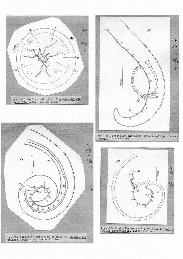
Fig. 27. Head end of male of Acanthocheilus quadridentatus. End-on view.
Fig. 28. Posterior extremity of male ot Rhabdochona amago. lateral view.
Fig. 29. Posterior extremity of male of Rhabdochona tridentigeris n. sp., lateral view.
Fig. 30. Posterior extremity of male of Hedruris neobythis, lateral view.
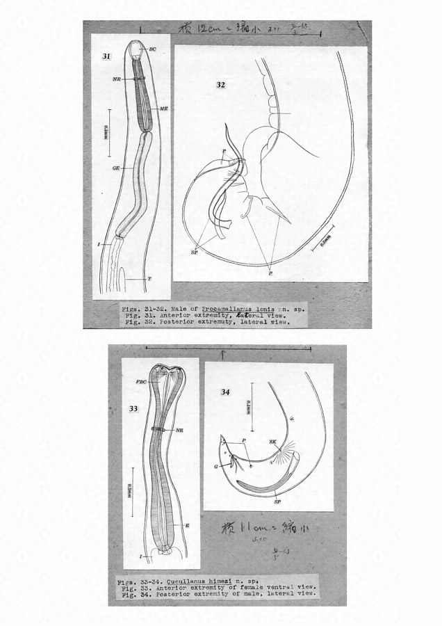
Figs. 31-32. Male ot Procamallanus lonis n. sp.: Fig. 31. Anterior extremity. lateral view., Fig. 32. Posterior extremity, lateral view.
Figs.33-34. Cucullanus himezi n. sp.: Fig. 33. Anterior extremity of female. ventral view., Fig. 34. Posterior extremity of male. lateral view.
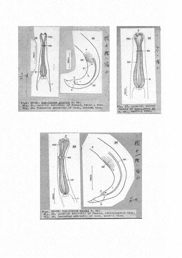
Figs. 35-36. Cucullanus cyprini n. sp.: Fig. 35. Anterior extremity of female, ventral view., Fig. 36. Posterior extremity of male, lateral view.
Fig. 37. Anterior extremity of female of Cucullanus girellae n. sp., ventral view.
Figs. 38-39. Cucullanus amadai n. sp.: Fig. 38. Anterior extremity of female, ventrolateral view., Fig. 39. Pasterior extremity of male, Lateral view.
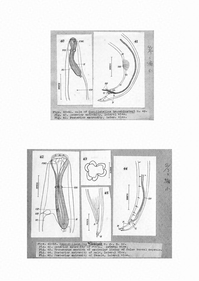
Figs. 40-41. Male of Cucullanellus branchiostegi n. sp.: Fig. 40. Anterior extremity, lateral view., Fig. 41. Posterior extremity, lateral view.
Figs. 42-45. Neocucullanellus apharei n. g., n. sp.: Fig. 42. Anterior extremity of male, lateral view., Fig. 43. Transverse section of cuticular lining of false buccal capsule., Fig. 44. Posterior extremity of male, lateral view., Fig. 45. Posterior extremity of female, lateral view.
所蔵館のウェブサイトで見る
公益財団法人 目黒寄生虫館文化庁 〒602-8959 京都府京都市上京区下長者町通新町西入藪之内町85番4
(C) The Agency for Cultural Affairs