寄生蠕虫類の分類体系.第Ⅲ巻.脊椎動物の線虫類 原図 5/10
きせいぜんちゅうるいのぶんるいたいけい.だいさんかん.せきついどうぶつのせんちゅうるい げんず じゅうぶんのご
概要
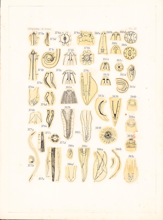
Plate 41.
Fig. 375. Aviculariella alcyona Wehr, 1931. a. Head, ventral view. b. Anterior extremity, lateral view. c. Posterior extremity of male. Gubernaculum.
Fig. 376. Trichostrongylus tenuis (Mehlis in Crepl., 1846). In Travassos, 1937. a. Bursa, lateral view. b. Dorsal and externodorsal rays. c—e. Spicules.
Fig. 377. Aproctella nuda Hamann, 1940. a. Female. b. Posterior extremity of male. c. Anterior extremity of female. d. Head, end-on view. e. Posterior extremity of female.
Fig. 378. Ancyracanthopsis fausti Li, 1934. a. Head, end-on view. b. Head, ventral view. c. Head, lateral view.
Fig. 379. Serticeps vulvoinflatum (Molin, 1860). After Drasche in Yorke and Maplestone, 1926. a. Head, lateral view. b. Head, end-on view. c. Posterior extremity of male.
Fig. 380. Paracuaria macdonaldi Rao, 1951. a. Anterior extremity. b. Head, lateral view. c. Head, ventral view. d—e. Head, end-on view at different levels.
Fig. 381. Habronema circi Li, 1934. a. Head, ventral view. b. Head, lateral view. c. Head, end-on view. d—e. Posterior extremity of male.
Fig. 382. a. Head of Ceratospira ophthalmica (Linstow, 1898). b. Posterior extremity of male of Ceratospira ophthalmica (Linstow, 1898). After Schneider. c. Posterior extremity of male of Ceratospira vesiculosa Schneider, 1866. After Linstow.
Fig. 383. Subulura forcipata (Rud., 1819). After Chow, 1939. a—b. Posterior extremity of male.
Fig. 384. Histiocephalus tridens Gendre, 1921. After Gendre in Y. & M., 1926. a. Head, ventral view. b. Head, partial end-on view.
Fig. 385. Histiocephalus laticaudatus (Rud., 1819). After Drasche in Yorke and Maplestone, 1926. a. Head, ventral view. b. Head, end-on view. c. Posterior extremity of male.
Fig. 386. Parhadjelia neglecta Lent et Freitas, 1939. a. Posterior extremity of male. b. Spicules. c. Posterior extremity of female. d. Tail of female. e. Egg.
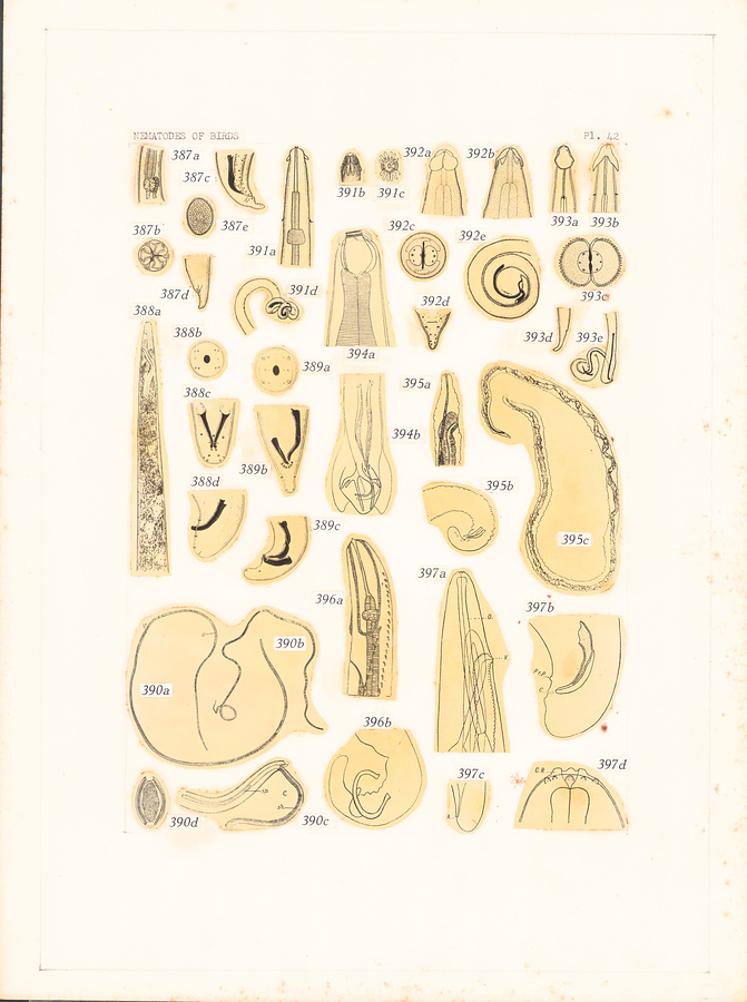
Plate 42.
Fig. 387. Porrocaecum wui Hsü, 1935. a. Esophago-intestinal region. b. Head, end-on view. c. Posterior extremity of male. d. Posterior extremity of female. e. Egg.
Fig. 388. Paramicipsella brevicaudata (Chow, 1939). a. Anterior extremity of female, lateral view. b. Head, end-on view. c—d. Posterior extremity of male.
Fig. 389. Paroncocerca tonkinensis Chow, 1939. a. Head, end-on view. b—c. Posterior extremity of male.
Fig. 390. Capillaria columbae (Rud., 1819). After Yorke and Maplestone, 1926. a. Female. b. Male. c. Posterior extremity of male. e. Egg.
Fig. 391. Skrjabinobronema coronatum (Molin, 1860). After Wehr, 1934. a. Anterior extremity, ventral view. b. Head, dorsal view. c. Head, end-on view. d. Posterior extremity of male.
Fig. 392. Schistorophus cucullatus Wehr, 1934. a. Head, ventral view. b. Head, lateral view. c. Head, end-on view. d. Posterior extremity of female. e. Posterior extremity of male.
Fig. 393. Stegophorus stellae-polaris (Parona, 1901). In Skrj. et al., 1949. a. Anterior extremity, lateral view. b. Anterior extremity, ventral view. c. Head, end-on view. d. Posterior extremity of female. e. Posterior extremity of male.
Fig. 394. Codiostomum struthionis Horst, 1885. After Y. ed M., 1926. a. Anterior extremity of male. b. Posterior extremity of male.
Fig. 395. Splendidofilaria pawlowskyi Skrj., 1923. After Skrj. in Yorke and Maplestone, 1926. a. Anterior extremity of female. b. Posterior extremity of male. c. Female.
Fig. 396. a. Anterior extremity of Echinuria Phoenicopteri (Seurat, 1916), lateral view. After Seurat in Y. & M., 1926. b. Posterior extremity of male of Echinuria jugadornata Soloview, 1912. After Soloview in Y. & M., 1926.
Fig. 397. Squamofilaria coronata (Rud., 1809). After Skrjabin, 1917. a. Anterior extremity of female, lateral view. b. Posterior extremity of male. c. Posterior extremity of female. d. Head, lateral view.
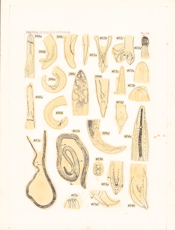
Plate 43.
Fig. 398. Paraprocta brevicauda (Chandler, 1924). After Maplestone, 1926. a. Anterior extremity of female. b. Posterior extremity of male. c. Posterior extremity of female.
Fig. 399. Dispharynx nasuta (Rud., 1819). After Yorke and Maplestone, 1926. a—b. Posterior extremity of male. c. Anterior extremity, ventral view. d. Posterior extremity of female.
Fig. 400. Pectinospirura argentata Wehr, 1933. a. Anterior extremity, lateral view. b. Posterior extremity of male.
Fig. 401. Trichosomoides crassicauda Bellingham, 1840. In Yorke and Maplestone, 1926. a. Mature female with male in uterus. b. Immature female with male in vagina.
Fig. 402. Ornithostrongylus nicobaricus Maplestone, 1940. a. Posterior extremity of female. b. Spicules and gubernaculum. c. Posterior extremity of male. d. Head, lateral view.
Fig. 403. Austroxyuris finlaysoni Johnston et Mawson, 1938. a-b. Head. c. Anterior extremity, ventral view. d. Posterior extremity of male.
Fig. 404. Gongylonema pulchrum Molin, 1857. After Baylis, 1939. a. Anterior extremity. b. Posterior extremity of male.
Fig. 405. Spiruracerca zapi Erickson, 1938. a. Posterior extremity of male. b. Head, end-on view. c. Head, lateral view. d. Posterior extremity of female.
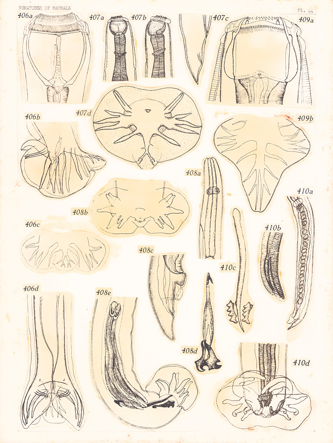
Plate 44.
Fig. 406. Alfortia edentata (Looss, 1900). After Skrjabin & Kutas, 1931. a. Head, dorsal view. b—c. Bursa. d. Posterior extremity of male.
Fig. 407. Castrostrongylus castoris Chapin, 1925. a. Anterior extremity, dorsal view. b. Anterior extremity, lateral view. c. Posterior extremity of female. d. Bursa.
Fig. 408. Capreocaulus capreoli (Stroh et Schmid, 1938). After Schulz and Kadenazii, 1948. a. Anterior extremity, lateral view. b. Bursa. c. Posterior extremity of female. d. Gubernaculum. e. Posterior extremity of male,
Fig. 409. Petrovinema skrjabini (Erschov, 1930) Erschov, 1943. a. Head. b. Bursa.
Fig. 410. Varestrongylus schulzi (Boev et Wolf, 1938). After Boev in Schulz and Boev, 1951. a. Posterior extremity of female. b. Distal end of spicule. c. Gubernaculum. d. Posterior extremity of male.
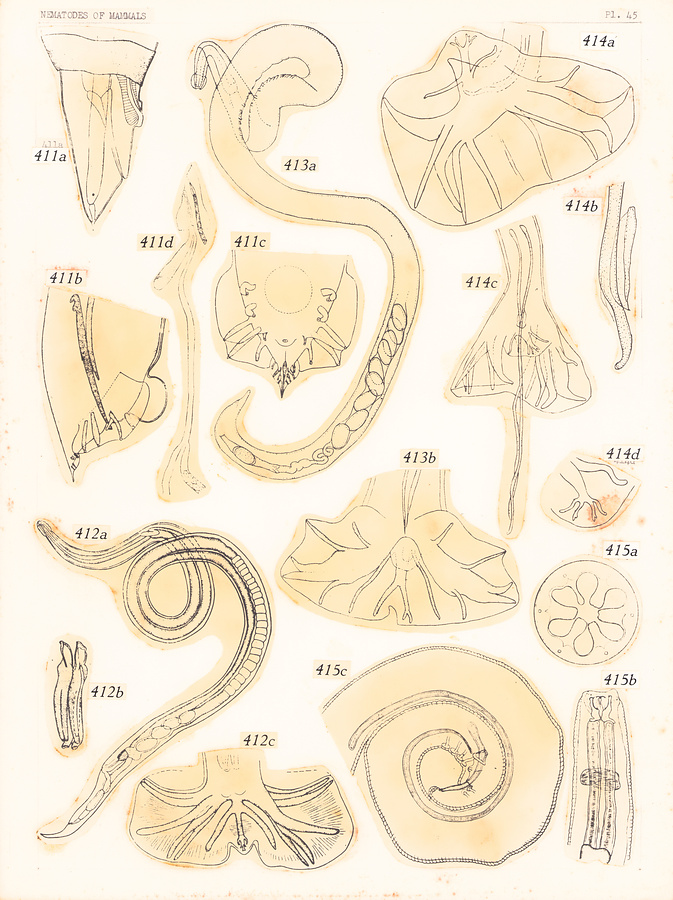
Plate 45.
Fig. 411. Maupasina weissi Seurat, 1913. After Seurat, 1917. a. Posterior extremity of female. b—c. Posterior extremity of male. d. Female terminalia.
Fig. 412. Mönningia mönningi Travassos, 1935. a. Female. b. Spicules. c. Posterior extremity of male.
Fig. 413. Acanthostrongylus acanthostrongylus Travassos, 1937. a. Female. b. Posterior extremity of male.
Fig. 414. Nippostrongylus braziliensis (Trav., 1914). After Travassos, 1937. a. Bursa. b. Spicule and gubernaculum. c. Posterior extremity of male. d. Dorsal ray.
Fig. 415. Petrowospirura lynxi Machulski, 1952. a. Head, end-on view. b. Anterior extremity. c. Posterior extremity of male.
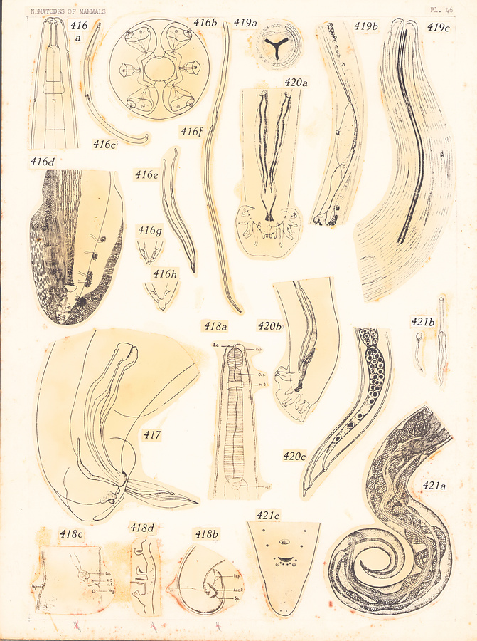
Plate 46.
Fig. 416. Didelphonema longispiculatum (Hill, 1939). After Hill, 1939. a. Anterior extremity, ventral view. b. Head, end-on view. c. Female. d. Posterior extremity of male. e—f. Spicules. g—h. Tail of female.
Fig. 417. Delamurella hyperoodoni Gubanov, 1952. posterior extremity of male.
Fig. 418. Monovaria rhinolophensia Khera, 1955. a. Anterior extremity of female. b. Posterior extremity of male. c. Vulvar region. d. Precloacal cuticular appendages.
Fig. 419. Pararhabdonema longistriatum Kreis, 1945. a. Transverse section of esophagus. b. Vulvar region. c. Anterior extremity.
Fig. 420. Parelaphostrongylus odocoilei (Hobmaier et Hobmaier, 1934). a—b. Posterior extremity of male. c. Posterior extremity of female.
Fig. 421. Cordophilus sagitta (Linst., 1907). After Mönnig, 1926. a. Anterior extremity of female. b. Spicules. c. Posterior extremity of male.
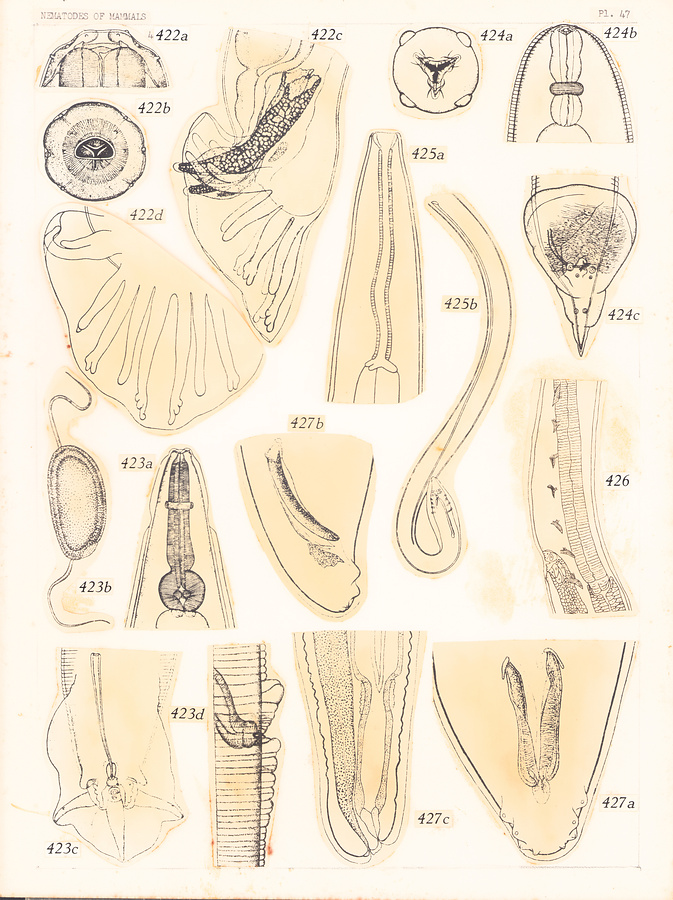
Plate 47.
Fig. 422. Dictyocaulus filaria (Rud., 1809). After Boev, 1952, in Skrjabin et al., 1952. a. Head, lateral view. b. Head, end-on view. c. Posterior extremity of male. d. Bursa.
Fig. 423. Citellina dispar Prendel, 1928. After Schulz, 1930. a. Anterior extremity. b. Egg. c. Posterior extremity of male. d. Vulva.
Fig. 424. Dentostomella translucida Schulz et Krepkogorskaja, 1932. a. Head, end-on view. b. Anterior extremity. c. Posterior extremity of male.
Fig. 425. Paraleiuris locchii Vaz et Pereira, 1929. a. Anterior extremity. b. Posterior extremity of male.
Fig. 426. Rictularia lucifugus Douvres. 1956; vulvar region.
Fig. 427. Anafilaroides rostrata Gerichter, 1949. a—b. Posterior extremity of male. c. Posterior extremity of female.
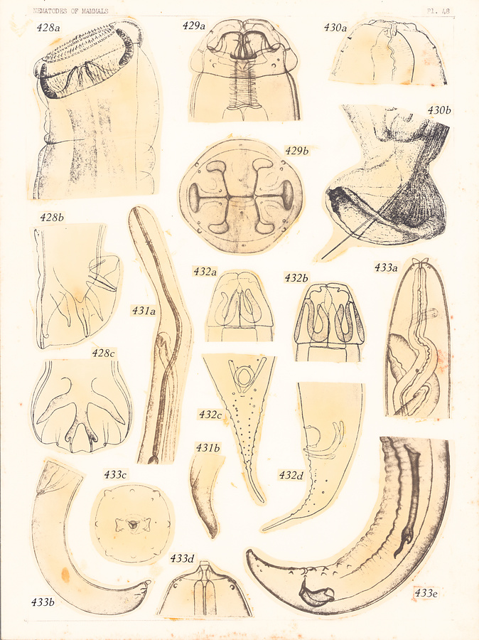
Plate 48.
Fig. 428. Okapistrongylus epuluensis van den Berghe, 1937. a. Anterior extremity. b—c. Bursa.
Fig. 429. Pygarginema skrjabini Kadenazii, 1948. a. Head, lateral view. b. Head, end-on view.
Fig. 430. Mirandonema intestinale Kreis, 1945. a. Head. b. Posterior extremity of male.
Fig. 431. Dipetalonema loxodontis (van den Berghe, 1937). a. Anterior extremity of female. b. Posterior extremity of female.
Fig. 432. Ansiruptodera ansirupta (Proença, 1937). a. Head, ventral view. b. Head, lateral view. c—d. Posterior extremity of male.
Fig. 433. Setaria kabargi Kadenazii, 1948. a. Anterior extremity of female. b. Posterior extremity of female. c. Head, end-on view. d. Head, lateral view. e. Posterior extremity of male.
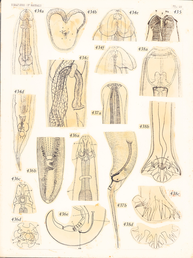
Plate 49.
Fig. 434. Cephaluris andrejevi Schulz, 1948. a. Anterior extremity, dorsal view. b. Head, end-on view. c. Vulvar region. d. Posterior extremity of male. e. Head, ventral view. f. Head, lateral view.
Fig. 435. Odontogeton phacochoeri Allgén, 1921; head, ventral view.
Fig. 436. Parabronema indicum Baylis, 1921. a. Anterior extremity, dorsal view. b. Posterior extremity of male. c. Head, lateral view. d. Head, end-on view. e. Posterior extremity of male.
Fig. 437. Carolodelatorrella tiflophila Vigueras, 1943. a. Head. b. Posterior extremity of male.
Fig. 438. Delafondia vulgaris (Looss, 1900). After Skrjabin and Kutas, 1931. a. Head, ventral view. b. Posterior extremity of male. c—d. Bursa.
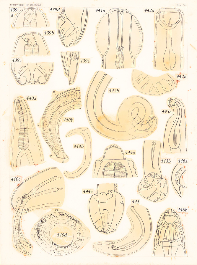
Plate 50.
Fig. 439. Galoncus perniciosus (Linstow, 1885). After Yorke and Maplestone, 1926. a. Head, dorsal view. b. Same, lateral view. c—d. Bursa. e. Posterior extremity of female.
Fig. 440. Filariopsis aspera van Thiel, 1926. a. Anterior extremity. b. Posterior extremity of female. c. Posterior extremity of male. d. Microfilaria.
Fig. 441. Mazzia mazzia Khalil et Vogelsang, 1932. a. Head. b. Posterior extremity of male.
Fig. 442. Mammomonogamus laryngeus (Raill., 1899). After Ryjikov, 1948, in Ryjikov, 1949. a. Anterior extremity. b. Bursal rays.
Fig. 443. Monodontus rarus Travassos, 1929. a. Anterior extremity. b. Posterior extremity of male.
Fig. 444. Pseudophysaloptera soricina Baylis, 1934. a. Head, lateral view. b. Female. c. Posterior extremity of male.
Fig. 445. Elaeophora böhmi Supperer, 1953; posterior extremity of male.
Fig. 446. Plicatolabia hagenbecki (Khalil et Vogelsang, 1932). a. Posterior extremity of male. b. Head.
所蔵館のウェブサイトで見る
公益財団法人 目黒寄生虫館文化庁 〒602-8959 京都府京都市上京区下長者町通新町西入藪之内町85番4
(C) The Agency for Cultural Affairs