寄生蠕虫類の分類体系.第Ⅳ巻.脊椎動物の単生類および盾吸虫類 原図 6/13
きせいぜんちゅうるいのぶんるいたいけい.だいよんかん.せきついどうぶつのたんせいるいおよびたてきゅうちゅうるい げんず じゅうさんぶんのろく
概要
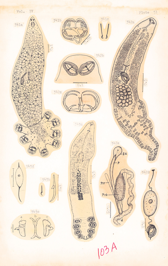
Plate 51.
Fig. 341a–c. Choricotyle hysteroncha (Fujii, 1944) (a), clamp (b), and posterior anchors (c).
Fig. 342a–e. Microcotyloides incisa (Linton, 1910) (a), oral suckers (b), clamp (c), egg (d), and male terminalia (e). After Fujii, 1944.
Fig. 343a–d. Hexostoma macracanthum Fujii, 1944 (a), posterior anchors (b), clamp skeleton of anteriormost (left) haptoral sucker (c), and egg (d).
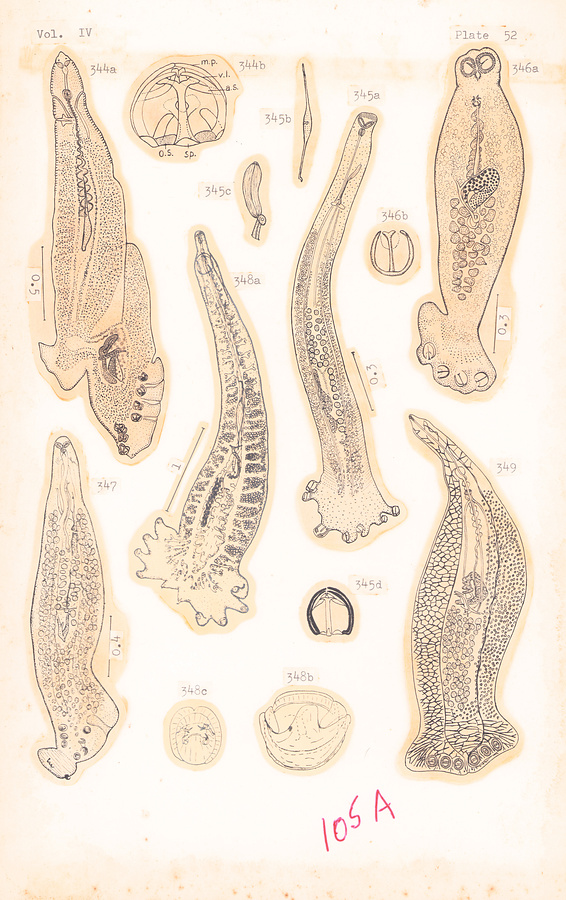
Plate 52.
Fig. 344a–b. Vallisiopsis contorta Subhapradha, 1951 (a) and clamp (b).
Fig. 345a–d. Gemmaecaputia corrugata Tripathi, 1959 (a), egg (b), cirrus (c), and clamp (d).
Fig. 346a–b. Hemilagia galapagensis (Meserve, 1958) (a) and clamp (b). After Meserve in Sproston, 1946.
Fig. 347. Bilaterocotyle chirocentrosus Chauhan, 1945.
Fig. 348a–c. Clupeocotyle brevoortia Hargis, 1955 (a), clamp (b) and genital atrim (c).
Fig. 349. Hexostoma grossum (Goto, 1894). Ca. 18 mm.
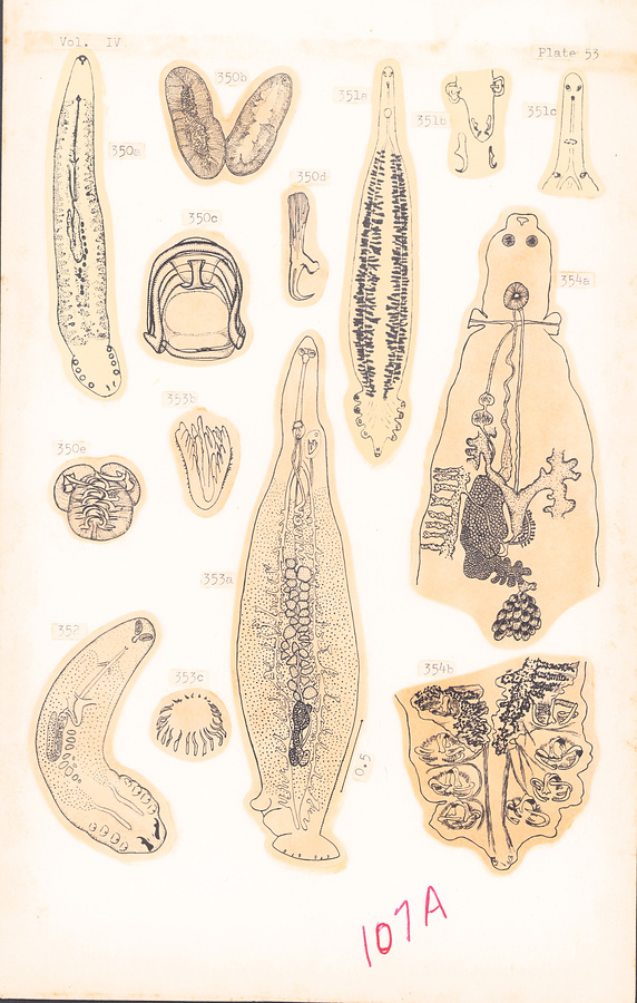
Plate 53.
Fig. 350a–e. Kuhnia scombri (Kuhn, 1829) (a) (×50), after Monticelli in Palombi, 1949; anterior suckers (b), clamp (c), terminal anchor (d) and genital hooks (e), in Sproston, 1946.
Fig. 351a–c. Winkenthughesia bramae (Parona et Perugia, 1896) (a) (×8), posterior extremity of body and hooks (b), and anterior extremity of body (c). After Parona and Perugia in Palombi, 1949.
Fig. 352. Kuhnia macracantha (Meserve, 1938).
Fig. 353a–c. Lethacotyle fijiensis Manter et Prince, 1953 (a), vagina (b) and genital crown of spines (c).
Fig. 354a–b. Winkenthugesia thyrsites (Hughes, 1928); anterior extremity (a), and posterior extremity (b).
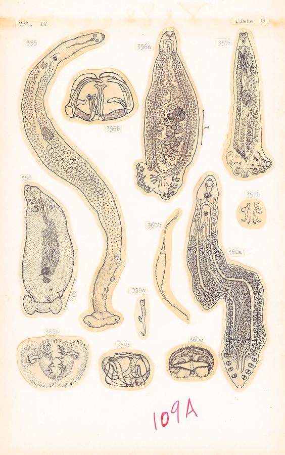
Plate 54.
Fig. 355. Protomicrocotyle mirabilis (MacCallum, 1918). Ovary actually posttesticular contrary to MacCallum's original figure!
Fig. 356a–b. Discocotyle sagittata (Leuckart, 1842) (a) after Bychowsky, 1957; clamp (b) after Sproston, 1946.
Fig. 357a–b. Plectanocotyle lorenzii Monticelli, 1899 (a) (×33), terminal anchors (b) (ca. ×166). After Monticelli in Palombi, 1949.
Fig. 358. Protomicrocotyle pacifica Meserve, 1938. After Meserve in Bychowsky, 1957.
Fig. 359a–c. Kuhnia minor (Goto, 1894); genital pore and hooks in dorsal view (a), clamp in dorsal view (b) and anchor (c). After Sproston, 1946.
Fig. 360a–c. Vallisia striata Parona et Perugia, 1890 (a) (×10), egg (b) (×30), clamp (c) (×100). After Monticelli, 1912, in Palombi, 1949.
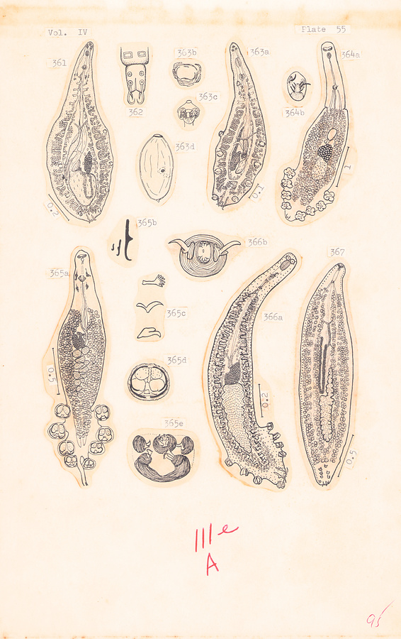
Plate 55.
Fig. 361. Mazocraeoides optisthonema Hargis, 1955.
Fig. 362. Ophicotyle fintae v. Beneden et Hesse, 1863.
Fig. 363a–d. Mazocraeoides georgei Price, 1936 (a), clamp (b), genital atrium (c), and egg (d). After Hargis, 1955.
Fig. 364a–b. Neomazocraes anadontostomae Tripathi, 1959 (a), and genital atrium (b).
Fig.365a–e. Paramazocraes thrissocles Tripathi, 1959 (a), terminal anchors (b), atrial hooks (c), clamp (d), and genital atrium (e).
Fig. 366a–b. Mazocraes gonialosae Tripathi, 1959 (a), and genital atrium (b).
Fig. 367. Kuhnia indica Tripathi, 1959.
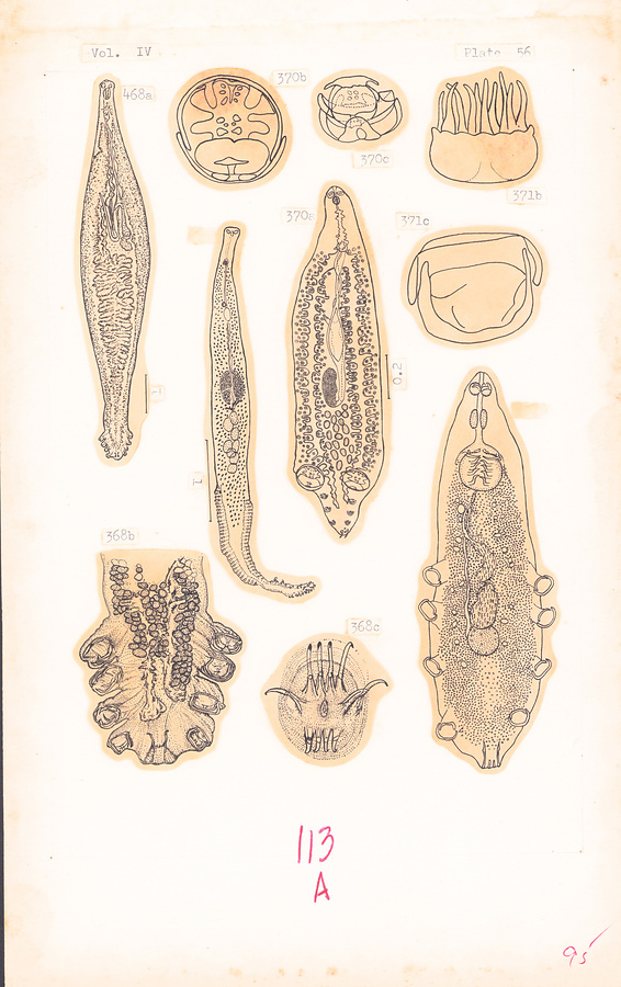
Plate 56.
Fig. 368a–c. Mazocraes alosae Hermann, 1782 (a), posterior extremity (b), and genital hooks (c). After Sproston, 1946.
Fig. 369. Microcotyle Pamae Tripathi, 1956.
Fig. 370a–c. Pseudoanthocotyle Pavlovskyi Bychowsky et Nagibina, 1954 (a), large clamp (b), and small clamp (c). After Bychowsky and Nagibina in Bychowsky, 1957.
Fig. 371a–c. Mazocraeoides prashadi Chauhan, 1950 (a) (0.5×0.14 mm), atrial spines (b), and clamp (c).
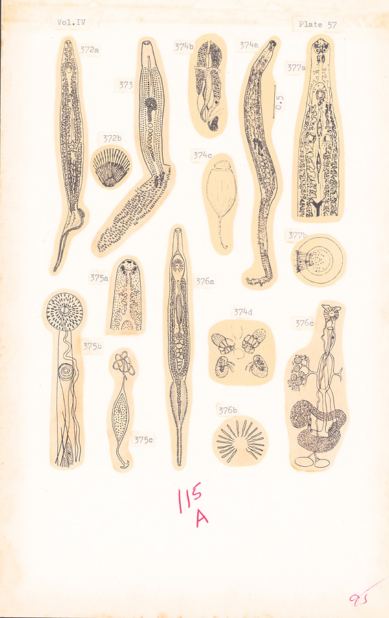
Plate 57.
Fig. 372a–b. Microcotyle chrysophryii v. Beneden et Hesse, 1863 in Parona et Perugia, 1890 (a) (×6) after Parona & Perugia in Palombi, 1949; atrial spines (b) (×130) after Palombi, 1949.
Fig. 373. Microcotyle priacanthi Meserve, 1938.
Fig. 374a–d. Cynoscionicola pseudoheteracantha (Hargis, 1957) (a), ovarian complex (b), egg (c), and genital atrium (d).
Fig. 375a–c. Microcotyle labracis v. Beneden et Hesse, 1863; anterior extremity (a), and region of genital pore and vagina (b) (ca. ×85) after Parona et Perugia in Palombi, 1949; and egg (c) (×86) after Palombi, 1949.
Fig. 376a–c. Microcotyle sargi Parona et Perugia, 1889 (a) (×15), genital crown of hooks (b), and genital complex (c). After Parona & Perugia in Palombi, 1949.
Fig. 377a–b. Bivagina alcedinis (Parona et Perugia, 1890) (a) (×45), and vaginal pore (b). After Parona & Perugia in Palombi, 1949.
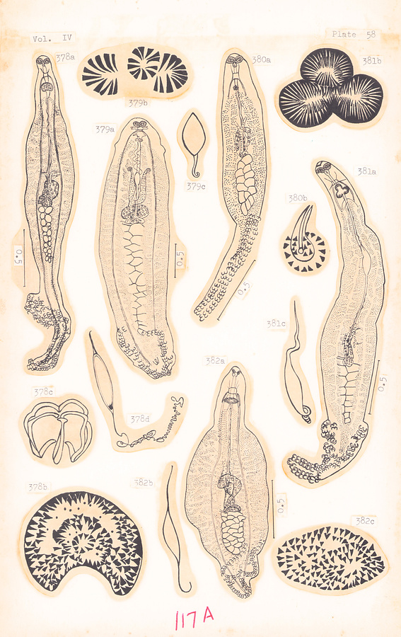
Plate 58.
Fig. 378a–d. Microcotyle temnodontis Sandars, 1944 (a); atrial spines (b), clamp (c), and egg (d).
Fig. 379a–c. Microcotyle agnostomi Sandars, 1945 (a); atrial spines (b), and egg (c).
Fig. 380a–b. Microcotyle parasillaginae Sandars, 1944 (a); cirrus and atrial spines (b).
Fig. 381a–c. Microcotyle odacis Sandars, 1945 (a); atrial spines (b), and egg (c).
Fig.382a–c. Microcotyle arripis Sandars, 1945 (a), egg (b), and atrial spines (c).
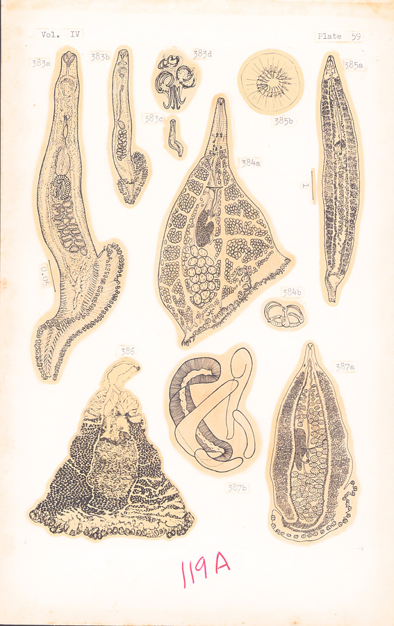
Plate 59.
Fig. 383a–d. Microcotyle donavini v. Benedcn et Hesse, 1863; adult with 46 pairs of clamps (a), very young adult with 26 pairs of clamps (b), larval form with 12 pairs of clamps (c), and posterior end of specimen in c (d). After Sproston, 1946.
Fig. 384a–b. Chlamydaxine truncata (Hargis, 1956) (a), and clamp (b).
Fig. 385a–b. Microcotyle Pseudomugilis Hargis, 1957 (a), and genital crown of hooks (b).
Fig. 386. Axine resplendens Caballero, Bravo Hollis et Grocott, 1954. 3.32 mm.
Fig. 387a–b. Allopseudaxine katsuwonis (Ishii, 1936) (a) after Ishii, 1936; clamp (b) original.
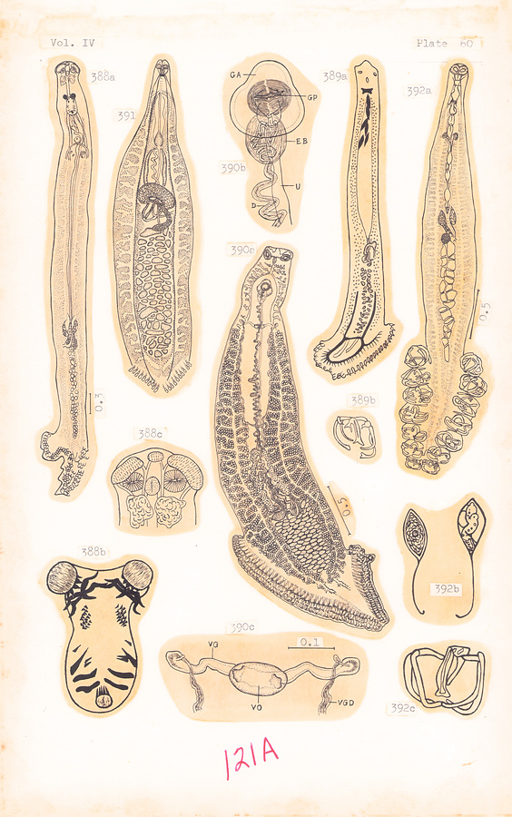
Plate 60.
Fig. 388a–c. Gonoplasius carangis Sandars, 1944 (a), genital atrium (b), and anterior extremity (c).
Fig. 389a–b. Cemocotylella elongata (Meserve, 1938) (a), and clamp (b).
Fig. 390a–c. Zeuxapta japonica nom. nov. for Microcotyle seriolae Yamaguti, 1940 (a), terminal genitalia (b), and vagina (c).
Fig. 391. Yamaguticotyla truncata (Goto, 1894). Ca. 3.3 mm.
Fig. 392a–c. Diplasiocotyle johnstoni Sandars, 1944 (a), eggs (b), and clamp (c).
所蔵館のウェブサイトで見る
公益財団法人 目黒寄生虫館文化庁 〒602-8959 京都府京都市上京区下長者町通新町西入藪之内町85番4
(C) The Agency for Cultural Affairs