寄生蠕虫類の分類体系.第Ⅳ巻.脊椎動物の単生類および盾吸虫類 原図 1/13
きせいぜんちゅうるいのぶんるいたいけい.だいよんかん.せきついどうぶつのたんせいるいおよびたてきゅうちゅうるい げんず じゅうさんぶんのいち
概要
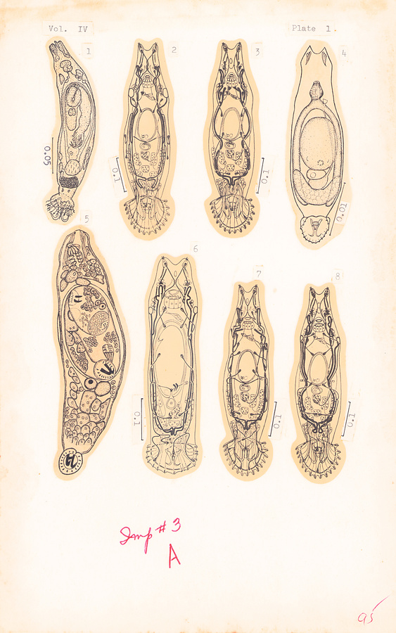
Plate 1. 1)
Fig. 1. Gyrodactylus funduli Hargis, 1955.
Fig. 2. Excretory system of Gyrodactylus flesi complex. After Malmberg, 1956.
Fig. 3. Excretory system of Gyrodactylus wageneri complex. After Malmberg, 1956.
Fig. 4. Gyrodactylus gobioninum Gussev, 1955.
Fig. 5. Metagyrodactylus indicus (Baugh, 1957).
Fig. 6. Excretory system of Gyrodactylus harengi complex. After Malmberg, 1956.
Fig. 7. Excretory system of Gyrodactylus elegans complex. After Malmberg, 1956.
Fig. 8. Excretory system of Gyrodactylus callariatis complex. After Malmberg, 1956.
1) Unless otherwise indicated all figures are from original sources. All scales are in mm, those without indication of length are 0.01 mm in value. It is impossible, however, to give scales or actual length for all figures reproduced herein because of relevant information lacking in some illustrations.
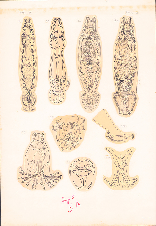
Plate 2.
Fig. 9. Macrogyrodactylus polypteri Malmberg, 1956.
Fig. 10. Gyrodactylus eucaliae Ikezaki et Hoffman, 1957.
Fig. 11. Gyrodactylus elegans Nordmann, 1832. After Wagener in Braun, 1893. ×240.
Fig. 12a–c. Macrogyrodactylus congolensis (Prudhoe, 1957) (a), opisthohaptor, lateral view (b), and anchor complex (c).
Fig. 13. Isancistrum loliginis de Beauchamp, 1912, in Bychowsky, 1957.
Fig. 14. Paragyrodactylus iliensis Gvozdev et Martekhov, 1953; anchor complex.
Fig. 15. Gyrodactyloides petruschewskii Bychowsky, 1947.
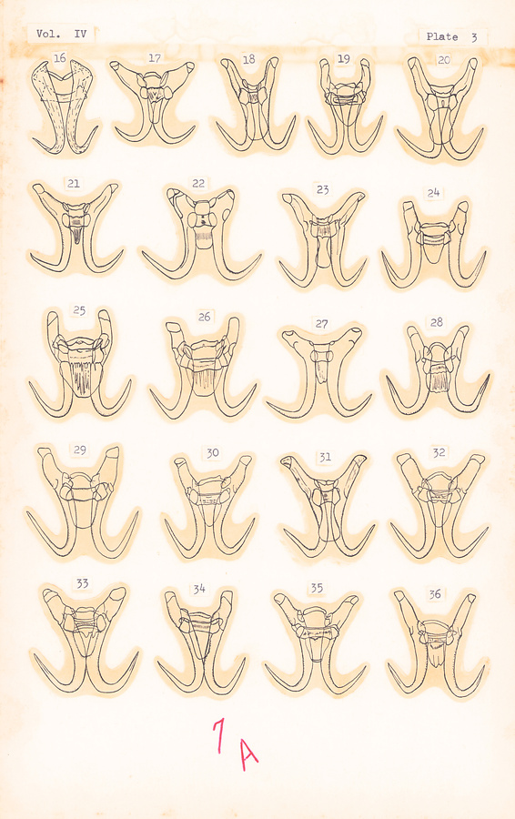
Plate 3.
(Anchor complex of Gyrodactylus species)
Fig. 16. G. funduli Hargis, 1955.
Fig. 17. G. harengi Malmberg, 1956.
Fig. 18. G. magnificus Malmberg, 1956.
Fig. 19. G. macronychus Malmberg, 1956.
Fig. 20. G. phoxini Malmberg, 1956.
Fig. 21. G. elegans of Malmberg, 1956.
Fig. 22. G. elegans minimus Malmberg, 1956.
Fig. 23. G. decorus Malmberg, 1956.
Fig. 24. G. wageneri lucii Malmberg, 1956.
Fig. 25. G. callariatis Malmberg, 1956.
Fig. 26. G. aculeati Malmberg, 1956.
Fig. 27. G. laevis Malmberg, 1956.
Fig. 28. G. robustus Malmberg, 1956.
Fig. 29. G. wageneri tincae Malmberg, 1956.
Fig. 30. G. wageneri aphyae Malmberg, 1956.
Fig. 31. G. carassii Malmberg, 1956.
Fig. 32. G. wageneri scardini Malmberg, 1956.
Fig. 33. G. salaris Malmberg, 1956.
Fig. 34. G. lavareti Malmberg, 1956.
Fig. 35. G. wageneri cernuae Malmberg, 1956.
Fig. 36. G. longiradix Malmberg, 1956.
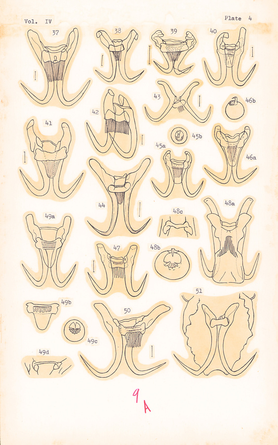
Plate 4.
(Anchor complex of Gyrodactylus species)
Fig. 37. G. barbatuli Achmerow, 1952.
Fig. 38. G. amurensis Achmerow, 1952.
Fig. 39. G. lefua Gussev, 1955.
Fig. 40. G. ophiocephali Gussev, 1955.
Fig. 41. G. rarus Wegener, 1909. After Gussev, 1955.
Fig. 42. G. oxycephali Achmerow, 1952.
Fig. 43. G. monstruosus Gussev, 1955.
Fig. 44. G. parvicopula Bychowsky, 1933. After Prost, 1957.
Fig. 45a–b. G. stephanus Mueller, 1937; anchors (a), and cirrus (b).
Fig. 46a–b. G. stegurus Mueller, 1937; anchors (a), and cirrus (b).
Fig. 47. G. gobioninum Gussev, 1955.
Fig. 48a–c. G. spathulatus Mueller, 1936; anchors (a), cirrus (b), and dorsal bar (c).
Fig. 49a–d. G. elegans muelleri Yin et Sproston, 1948; anchors (a), ventral bar (b), cirrus (c), and dorsal bar (d). After Mueller, 1936.
Fig. 50. G. curiosus Gussev, 1955.
Fig. 51. G. menschikowi Gvozdev, 1950.
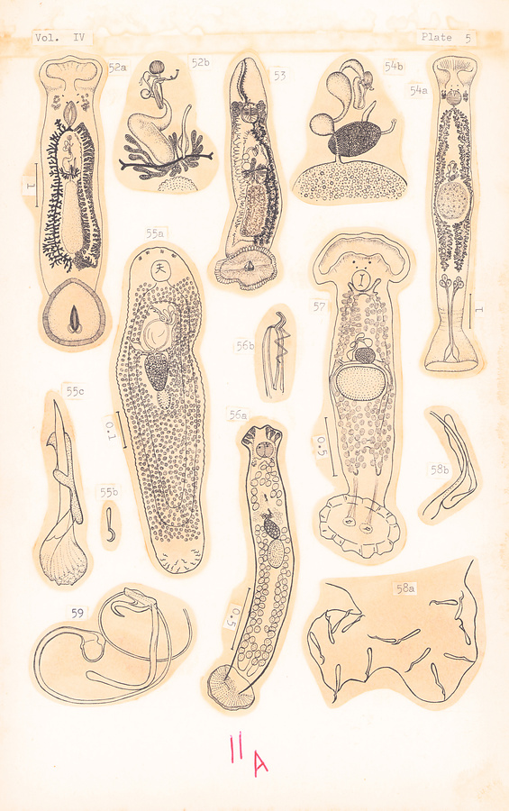
Plate 5.
Fig. 52a–b. Calceostoma calceostoma (Wagener, 1857) (a), and genital complex (b). After Palombi, 1949.
Fig. 53. Calceostoma glandulosum Johnston et Tiegs, 1922. Ca. 5 mm.
Fig. 54a–b. Calceostomella inermis (Parona et Perugia, 1889) (a), and genital complex (b). After Palombi, 1949.
Fig. 55a–b. Acolpenteron nephriticum Gvozdev, 1945 (a) after Bychowsky, 1957; haptoral hooks (b) and copulatory organ (c) after Gvozdev, 1945.
Fig. 56a–b. Neocalceostoma elongatum Tripathi, 1959 (a) and copulatory organ (b).
Fig. 57. Calceostomella inermis (Parona et Perugia). After Bychowsky, 1957.
Fig. 58a–b. Acolpenteron pavlowskii Bychowsky et Gussev, 1955 (a) and copulatory organ (b).
Fig. 59. Acolpenteron ignotum Gussev, 1955; copulatory organ.
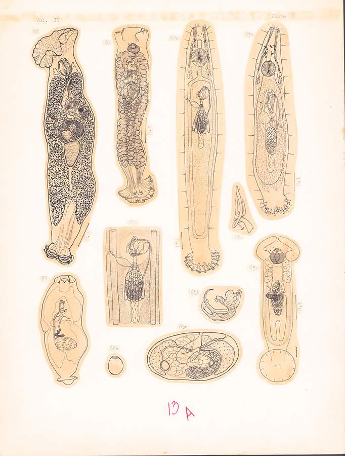
Plate 6.
Fig. 60. Paracalceostoma calceostomoides Caballero et Bravo Hollis, 1959.
Fig. 61. Pseudocalceostoma sciaenae Yamaguti, 1940.
Fig. 62a–c. Acolpenteron ureteroecetes Fischthal et Allison, 1940 (a), genital complex (b), and egg (c). After Fischthal and Allison, 1941.
Fig. 63a–b. Acolpenteron catostomi Fischthal et Allison, 1942, (a), and copulatory organ (b).
Fig. 64. Fridericianella ovicola Brandes, 1894. After Brandes in St. Remy, 1898.
Fig. 65a–c. Anoncohaptor anomalus Mueller, 1938, in Bychowsky, 1957 (a), copulatory apparatus (b), and transverse section through ovarian complex (c).
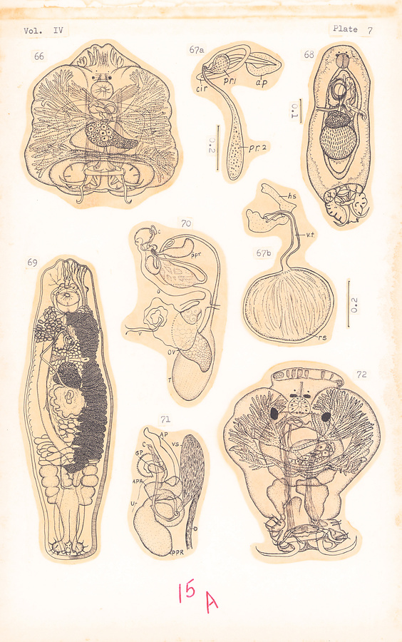
Plate 7.
Fig. 66. Protogyrodactylus quadratus Johnston et Tiegs, 1922. Ca. 0.23 mm.
Fig. 67a–b. Dactylogyrus orientalis Jain, 1959; copulatory organ (a), and vaginal complex (b).
Fig. 68. Dactylogyrus geei Yin et Sproston, 1949.
Fig. 69. Microncotrematoides inversum (Goto et Kikuchi, 1917), semidiagrammatic.
Fig. 70. Dactylogyrus robustus Malewitzkaja, 1941; genital complex. After Gussev, 1955.
Fig. 71. Dactylogyrus apogonis Yamaguti, 1940; genital complex.
Fig. 72. Trivitellina subrotunda Johnston et Tiegs, 1922. Ca. 0.2 mm.
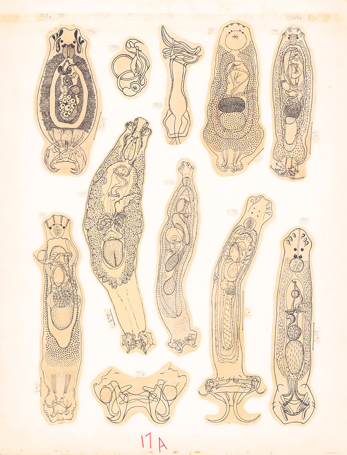
Plate 8.
Fig. 73a–b. Cichlidogyrus arthracanthus Paperna, 1960 (a) and copulatory organ (b).
Fig. 74. Protancyrocephalus strelkowi Bychowsky, 1957.
Fig. 75. Neodactylogyrus curvicirrus (Achmerow, 1952), in Gussev, 1955.
Fig. 76. Ancylodiscoides magnus Bychowsky et Nagibina, 1958.
Fig. 77. Mexicana bychowskyi Caballero et Bravo Hollis, 1959.
Fig. 78a–c. Ancyrocephalus platycephali (Yin et Sproston, 1948) (a), copulatory organ (b), and haptor (c).
Fig. 79. Mizelleus indicus Jain, 1957.
Fig. 80. Ancyrocephalus manilensis Tubangui, 1931.
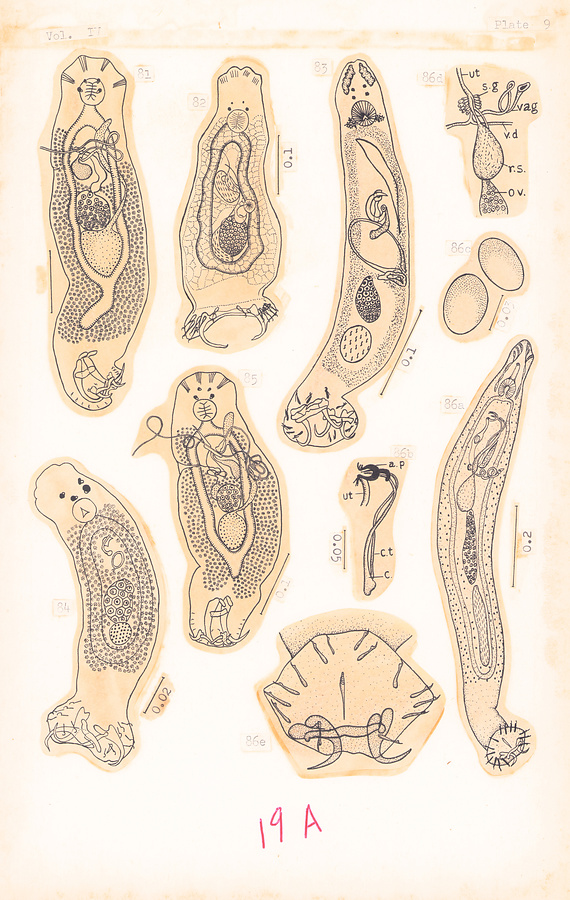
Plate 9.
Fig. 81. Ancylodiscoides siluri (Zandt, 1924). After Bychowsky, 1957.
Fig. 82. Dogielius forceps Bychowsky, 1936. After Bychowsky, 1957.
Fig. 83. Ancylodiscoides notopterus (Jain, 1955).
Fig. 84. Heteroncocleidus buschkieli Bychowsky, 1957.
Fig. 85. Ancylodiscoides vistulensis (Siwak, 1932). After Bychowsky and Nagibina, 1957.
Fig. 86a–e. Paradactylogyrus catlaius Thapar, 1948 (a), copulatory organ (b), eggs (c), genital complex (d), and opisthohaptor (e).
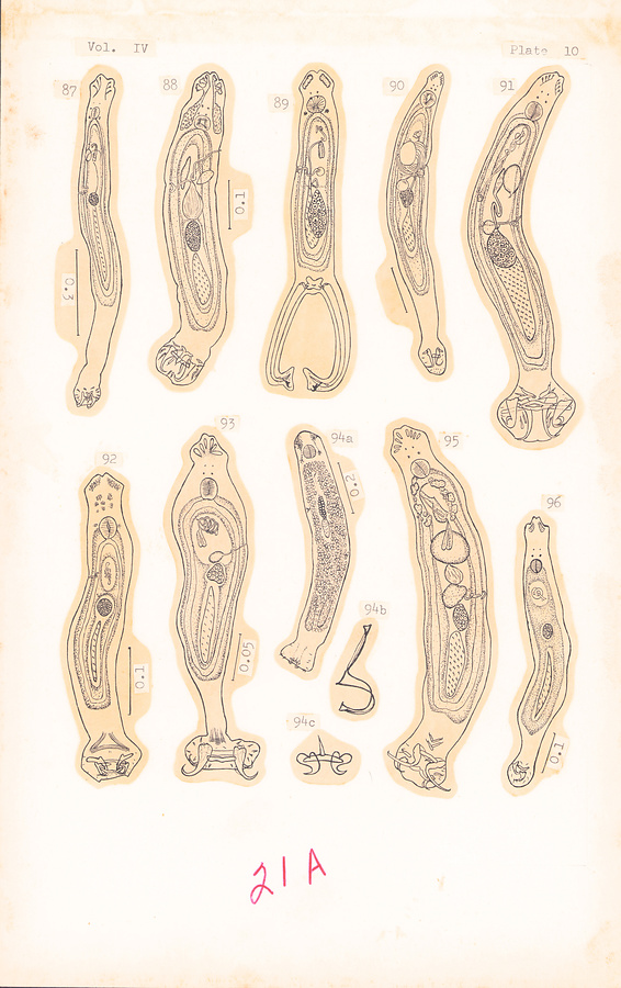
Plate 10.
Fig. 87. Neodactylogyrus indicus Jain, 1957.
Fig. 88. Neodactylogyrus calbasi Jain, 1957.
Fig. 89. Bifurcohaptor indicus Jain, 1958.
Fig. 90. Dactylogyrus multispiralis Jain, 1957.
Fig. 91. Neosprostonia indica Jain, 1959 (dorsal).
Fig. 92. Haplocleidus rhyncobdelli (Jain, 1959).
Fig. 93. Haplocleidus xenentodoni (Jain, 1959).
Fig. 94a–c. Paradactylogyrus bati Tripathi, 1959 (a), copulatory organ (b), and anchor complex (c).
Fig. 95. Neosprostonia wallagonia Jain, 1959 (dorsal).
Fig. 96. Neodactylogyrus cotius Jain, 1957.
所蔵館のウェブサイトで見る
公益財団法人 目黒寄生虫館文化庁 〒602-8959 京都府京都市上京区下長者町通新町西入藪之内町85番4
(C) The Agency for Cultural Affairs