寄生蠕虫類の分類体系.第Ⅳ巻.脊椎動物の単生類および盾吸虫類 原図 3/13
きせいぜんちゅうるいのぶんるいたいけい.だいよんかん.せきついどうぶつのたんせいるいおよびたてきゅうちゅうるい げんず じゅうさんぶんのさん
概要
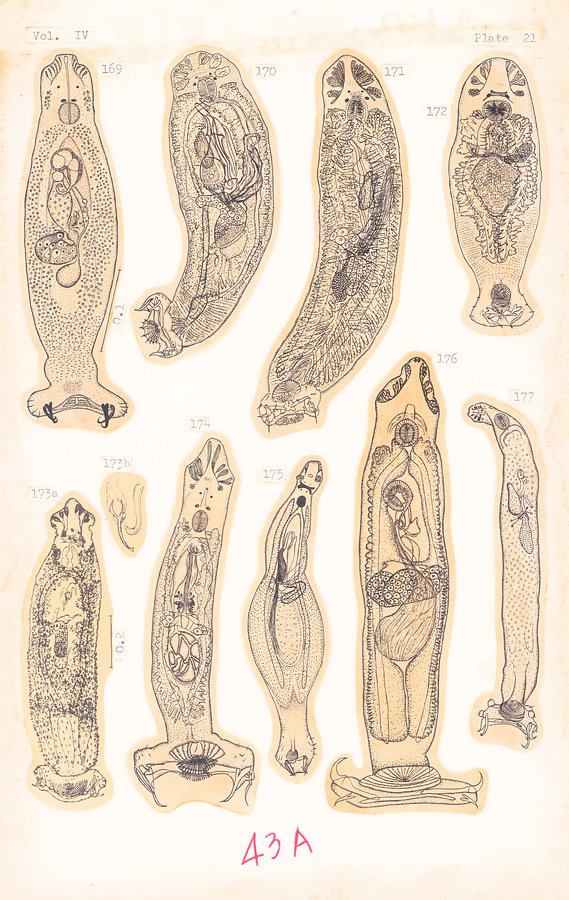
Plate 21.
Fig. 169. Diplectanum melanesiense Laird, 1958.
Fig. 170. Lepidotrema therapon Johnston et Tiegs, 1922. 0.5–0.77 mm.
Fig. 171. Lepidotrema tenue Johnston et Tiegs, 1922. 0.69 mm.
Fig. 172. Lepidotes fluviatilis Johnston et Tiegs, 1922. 0.95 mm.
Fig. 173a–b. Diplectanum bilobatum Hargis, 1955 (a), and copulatory apparatus (b).
Fig. 174. Lepidotrema angustum (Johnston et Tiegs, 1922). Ca. 0.32 mm.
Fig. 175. Lamellodiscus typicus Johnston et Tiegs, 1922. Ca. 0.124 mm.
Fig. 176. Acleotrema girellae Johnston et Tiegs, 1922. Ca. 0.7 mm.
Fig. 177. Lamellodiscoides belengeri (Chauhan, 1945).
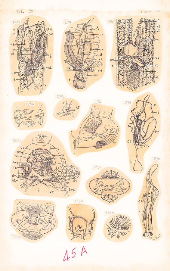
Plate 22.
Fig. 178a–c. Lepidotrema fuliginosum Johnston et Tiegs, 1922; genital complex (a), opisthohaptor (b), and female genital ducts (c).
Fig. 179. Lepidotrema tenue Johnston et Tiegs, 1922; genital complex.
Fig. 180. Lepidotrema simplex Johnston et Tiegs, 1922; genital complex.
Fig. 181. Lepidotrema angustum Johnston et Tiegs, 1922; opisthohaptor.
Fig. 182a–b. Lepidotes fluviatilis Johnston et Tiegs, 1922; genital complex (a) and opisthohaptor (b).
Fig. 183a–e. Lepidotrema therapon Johnston et Tiegs, 1922; opisthohaptor in end-on view (a), same in lateral view (b), squamodisc (c), entire male genital complex (d), and male terminalia (e).
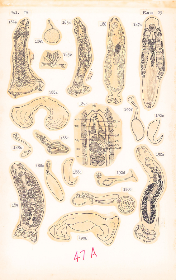
Plate 23.
Fig. 184a–b. Rhabdosynochus rhabdosynochus Mizelle et Blatz, 1941 (a), and egg (b). After Hargis, 1955.
Fig. 185a–b. Rhamnocercus bairdiella Hargis, 1955 (a), and accessory haptoral plaque (b).
Fig. 186. Salmoncus variabilis (Mizelle et Webb, 1953).
Fig. 187a–b. Linguadactyla molvae Brinkmann, 1940 (a) (×24), genital complex (b).
Fig. 188a–e. Tetraoncoides japonicus Bychowsky, 1951; bar (a), vagina (b), copulatory organ (c), anchor (d), and egg (e).
Fig. 189. Tetraoncus monenteron (Wagener, 1857), ×90. After Wegener, 1910.
Fig. 190a–f. Tetraoncoides paradoxus Bychowsky, 1951 (a), bar (b), copulatory organ (c), vagina (d), anchor (e), and egg (f).
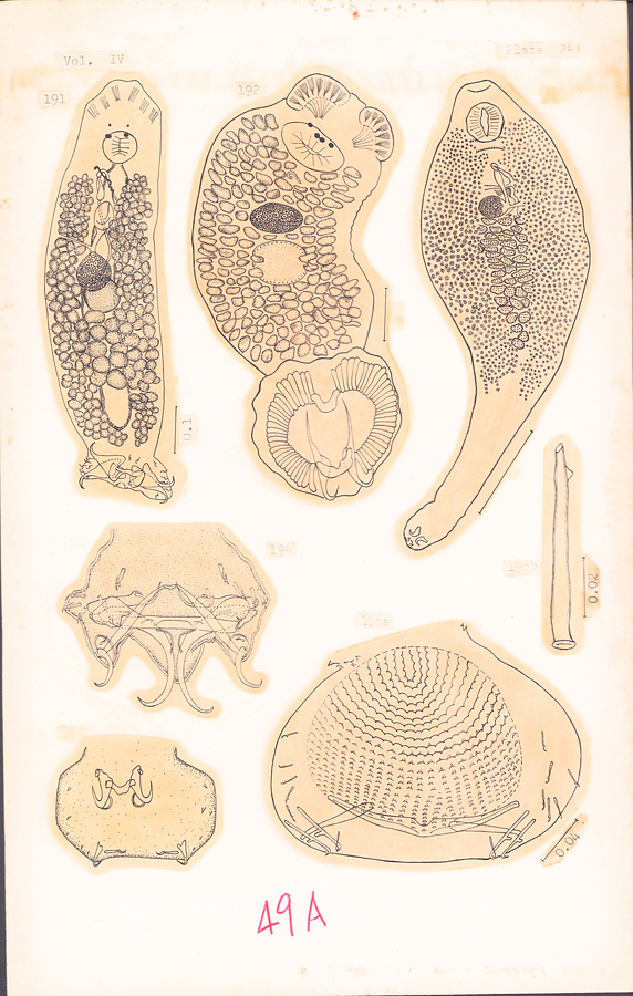
Plate 24.
Fig. 191. Tetraoncus monenteron (Wagener, 1857). After Bychowsky, 1957.
Fig. 192. Bothitrema bothi (MacCallum, 1913). After Bychowsky, 1957.
Fig. 193. Linguadactyla molvae Brinkmann, 1940. After Bychowsky, 1957.
Fig. 194. Mizelleus indicus Jain, 1957; opisthohaptor.
Fig. 195. Thaparocleidus wallagonius Jain, 1952; opisthohaptor.
Fig. 196a–b. Diplectanum collinsi (Mueller, 1936); opisthohaptor (a) and cirrus (b).
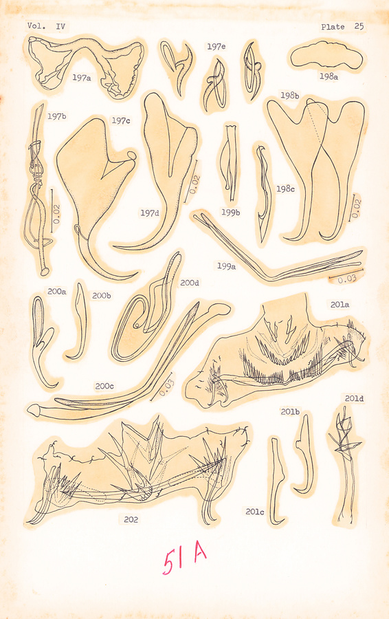
Plate 25.
Fig. 197a–e. Tetraoncus monenteron (Wagener, 1857); dorsal bar (a), copulatory organ (b), ventral anchor (c), dorsal anchor (d), and marginal hooklets (e). After Prost, 1957.
Fig. 198a–c. Salmoncus lenoki (Achmerow, 1952); bar (a), anchors (b), and copulatory organ (c).
Fig. 199a–b. Neodiplectanum wenningeri Mizelle et Blatz, 1941; dorsal and ventral bars (a) and copulatory organ (b).
Fig. 200a–d. Rhabdosynochus rhabdosynochus Mizelle et Blatz, 1941; ventral anchor (a), dorsal anchor (b), dorsal and ventral bars (c), and copulatory organ (d).
Fig. 201a–d. Rhamnocercus stichospinus Seamster et Monaco, 1956; opisthohaptor (a), dorsal anchor (b), ventral anchor (c), and copulatory organ (d).
Fig. 202. Rhamnocercus rhamnocercus Monaco, Wood et Mizelle, 1954; opisthohaptor.
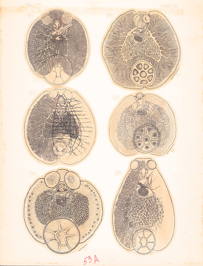
Plate 26.
Fig. 203. Capsala ovalis (Goto, 1894). Ca. 13 mm.
Fig. 204. Tristoma coccineum Cuvier, 1817. After Bychowsky, 1957.
Fig. 205. Capsaloides sinuata (Goto, 1894). Ca. 8 mm.
Fig. 206. Capsala martinierei Bosc, 1811. After Price, 1939.
Fig. 207. Capsala pricei Hidalgo Escalente, 1959. After Price, 1960.
Fig. 208. Capsala nozawae (Goto, 1894).
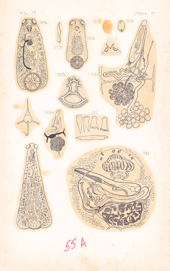
Plate 27.
Fig. 209a–c. Capsala pelamydis (Taschenberg, 1878) (a) (×13), genital complex (b), egg (c). After Parona & Perugia in Palombi, 1949.
Fig. 210a–c. Capsala interrupta (Monticelli, 1891 (a) (×15), posterior part of opisthohaptor (b) (×39), and anchor (c). After Monticelli in Palombi, 1949.
Fig. 211a–d. Tristoma coccineum Cuvier, 1817; dorsal view (a) (×1/8), ventral view (b), frontal plate extended into two horns (c), genital complex (d) (×16). After Taschenberg in Palombi, 1949.
Fig. 212. Tristoma coccineum Cuvier, 1817; dorsal marginal spines and anchor. After Price, 1939.
Fig. 213. Capsaloides perugiai (Setti, 1898), ×15. After Setti in Palombi, 1949.
Fig. 214. Capsala pricei Hidalgo Escalente, 1959; pharynx and genital complex.
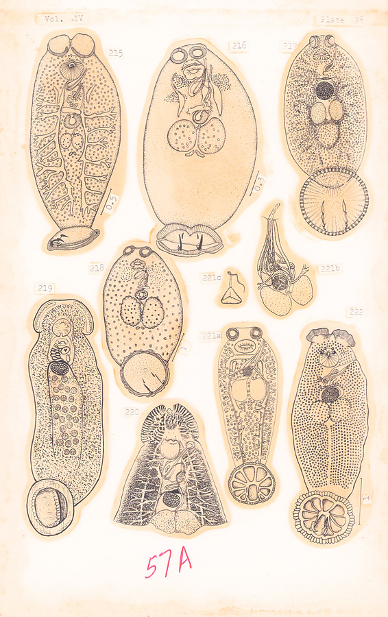
Plate 28.
Fig. 215. Neobenedenia muelleri (Meserve, 1938).
Fig. 216. Neobenedenia adenea (Meserve, 1936). After Meserve, 1938.
Fig. 217. Benedenia monticellii (Parona et Perugia, 1895), 1.75 mm. After Palombi, 1949.
Fig. 218. Neobenedenia isabellae (Meserve, 1938).
Fig. 219. Nitzschia sturionis (Abildgaard, 1794). After Brinkmann, 1952.
Fig. 220. Entobdella diadema (Monticelli, 1902), ×12. After Monticelli in Palombi, 1949.
Fig. 221a–c. Trochopus heteracanthus Massa, 1903 (a) (×16), genital complex (b), and egg (c). After Massa in Palombi, 1949.
Fig. 222. Trochopus pini (v. Beneden et Hesse, 1863). After Bychowsky, 1957
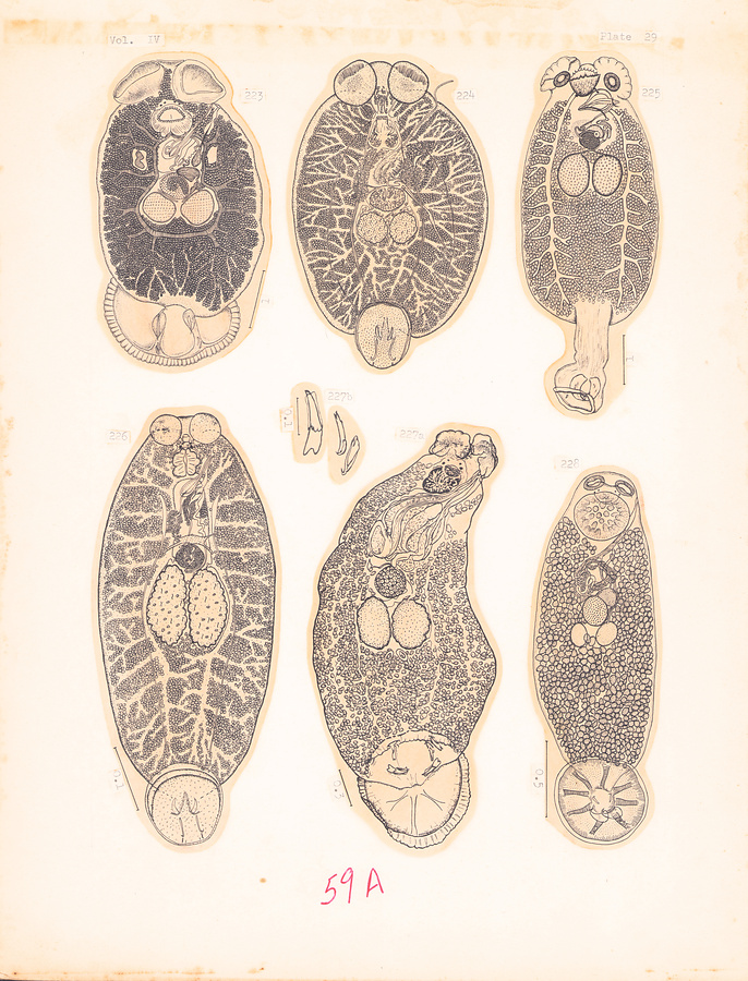
Plate 29.
Fig. 223. Benedenia derzhavini (Layman, 1930). After Bychowsky, 1957.
Fig. 224. Benedeniella posterocolpa (Hargis, 1955).
Fig. 225. Encotyllabe spari Yamaguti, 1934.
Fig. 226. Neobenedenia girellae (Hargis, 1955).
Fig. 227a–b. Trochopus pseudomarginatus Bravo Hollis, 1958 (a) and three pairs of anchors (b).
Fig. 228. Trochopus goniistii Yamaguti, 1940.
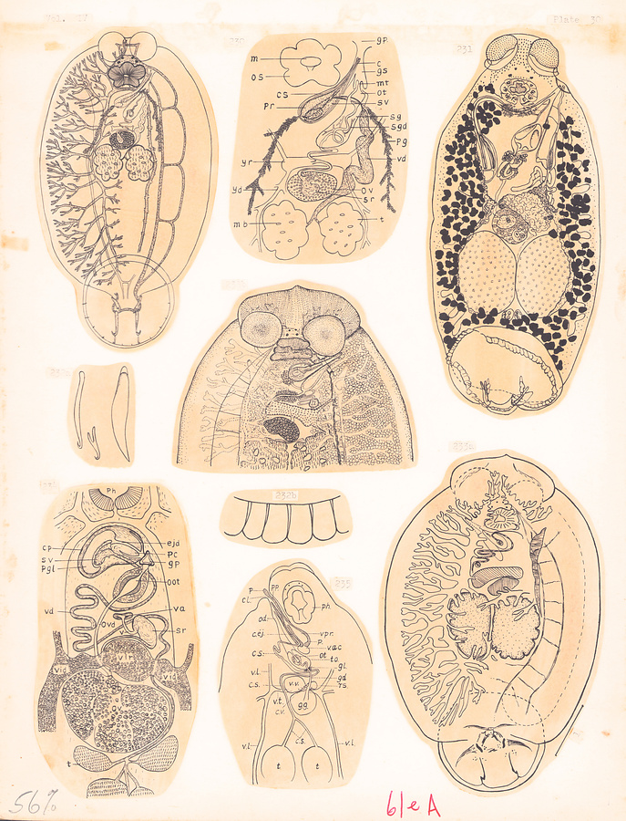
Plate 30.
Fig. 229. Neobenedenia melleni (MacCallum, 1927). After Jahn and Kuhn, 1932.
Fig. 230. Neobenedenia melleni (MacCallum, 1927); genital complex. After Jahn and Kuhn, 1932.
Fig. 231. Pseudobenedenia noblei (Menzies, 1946).
Fig. 232a–b. Benedenia monticellii (Parona et Perugia, 1895); three pairs of anchors (a), marginal membrane of opisthohaptor (b). After Palombi, 1949.
Fig. 233a–b. Pseudobenedenia nototheniae Johnston, 1931 (a), and anterior extremity (b). After Johnston in Sproston, 1946.
Fig. 234. Nitzschia sturionis (Abildgaard, 1794); genital complex. After Brinkmann, 1952.
Fig. 235. Entobdella soleae (v. Beneden et Hesse, 1863); genital complex. After Saint Remy, 1891.
所蔵館のウェブサイトで見る
公益財団法人 目黒寄生虫館文化庁 〒602-8959 京都府京都市上京区下長者町通新町西入藪之内町85番4
(C) The Agency for Cultural Affairs