ハワイ産魚類の二生吸虫類 原図 3/9
はわいさんぎょるいのにせいきゅうちゅうるい げんず きゅうぶんのいち
概要
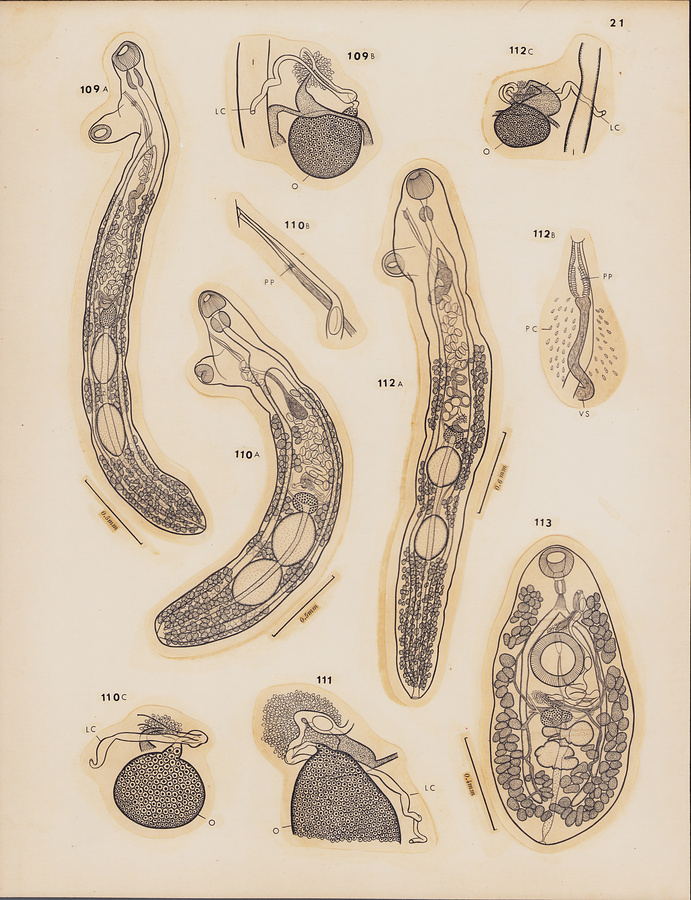
Plate 21
Fig.109 Pseudopecoeloides opelu n. sp. A, holotype, dorsal view; B, ovarian complex of paratype, dorsal view.
Fig.110 Pseudopecoeloides parviacetabulatus n. sp. A, holotype, lateral view anteriorly, dorsal view posteriorly; B, terminal genitalia of holotype; C, ovarian complex of holotype, dorsal view.
Fig.111 Pseudopecoeloides tenuoides Martin, 1960, ovarian complex of a specimen from Priacanthus cruentatus, ventral view.
Fig.112 Pseudopecoeloides wekeula n. sp. A, holotype, dorsal view; B, terminal genitalia of paratype, ventral view; C, ovarian complex of same, ventral view.
Fig.113 Pseudopecoelus acanthuri n. sp., holotype, ventral view.
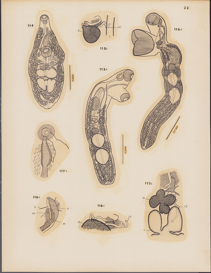
Plate 22
Fig.114 Pseudopecoelus puhipaka n. sp., holotype, ventral view.
Fig.115 Pseudopecoelus maomao n. sp. A, holotype, ventral view; B, ovarian complex of paratype, ventral view.
Fig.116 Pseudopecoelus sphyraenae n. sp. A, holotype from Sphyraena barracuda, lateral view anteriorly, ventral view posteriorly; B, terminal genitalia of holotype; C, ovarian complex of holotype, ventral view.
Fig.117 Pseudopecoelus vitellozonatus Pritchard, 1966. A, terminal genitalia of a specimen from Holocentrus spinifer; B, ovarian complex of a specimen from Holocentrus lacteoguttatus, ventral view.
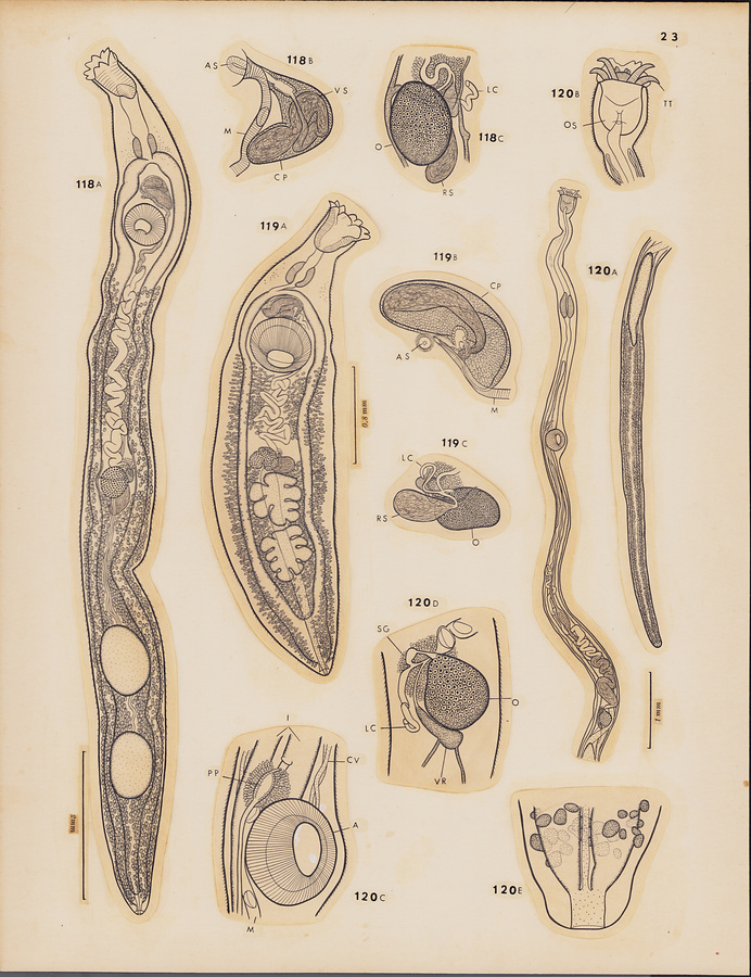
Plate 23
Fig.118 Enenterum elongatum n. sp. A, holotype, ventral view; B, terminal genitalia of paratype, ventral view; C, ovarian complex of paratype, ventral view.
Fig.119 Enenterum kyphosi n. sp. A, holotype, ventral view; B, terminal genitalia of paratype, ventral view; C, ovarian complex of paratype, dorsal view.
Fig.120 Jeancadenatia pacifica n. sp. A, holotype, ventral view; B, anterior extremity of holotype, ventral view; C, terminal genitalia of holotype, ventral view; D, ovarian complex of holotype, ventral view; E, posterior extremity of holotype, ventral view.
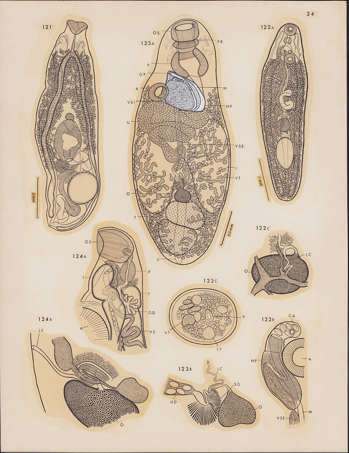
Plate 24
Fig.121 Bivesicula congeri n. sp., holotype, ventral view.
Fig.122 Hapladena spinosa Manter et Pritchard, 1961. A, entire worm from Acanthurus dussumieri, ventral view; B, terminal genitalia, ventral view; C, ovarian complex, dorsal view.
Fig.123 Metamegasolena scarideae n. g., n. sp. A, holotype, ventral view; B, ovarian complex, dorsal view; C, transverse section of paratype through posterior extremity, showing lymph vessels.
Fig.124 Tetrochetus aluterae (Hanson, 1955). A, forebody, lateral view; B, ovarian complex, lateral view.
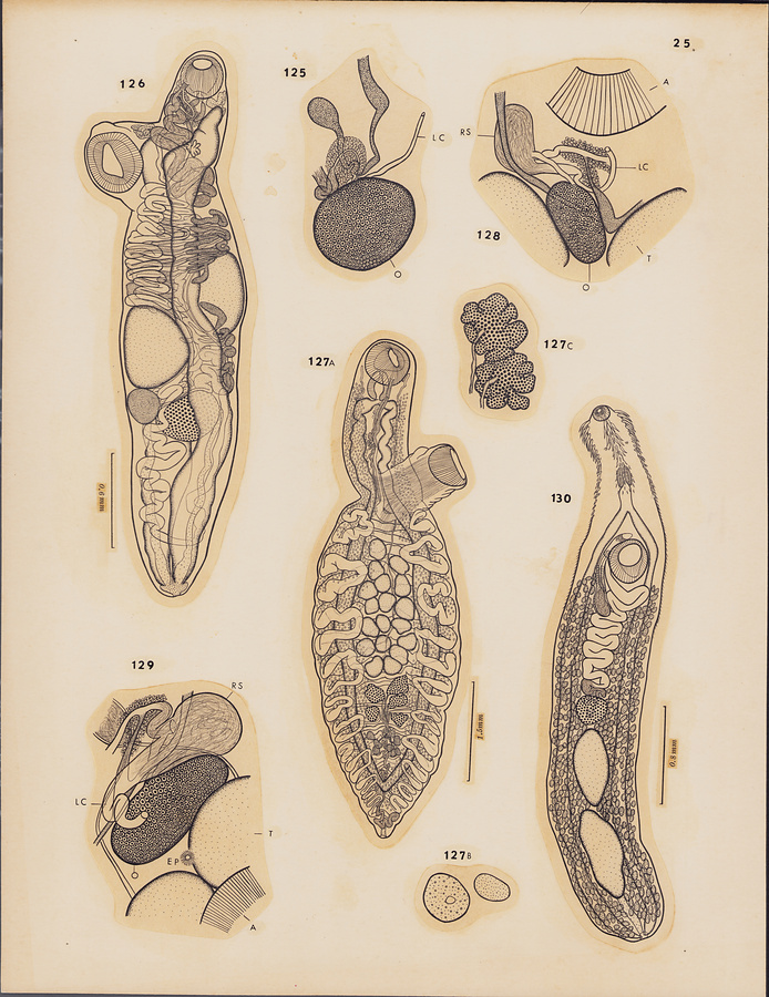
Plate 25
Fig.125 Tetrochetus coryphaenae Yamaguti, 1934, ovarian complex, dorsal view.
Fig.126 Tetrochetus macrorchis n. sp., holotype from Xanthichthys ringens, ventrolateral view.
Fig.127 Syncoelium cypseluri n. sp. A, holotype, ventral view; B, ""Drüsenartige Zellennester"" of Looss; C, two ovaries of paratype, ventral view.
Fig.128 Heterolebes maculosus Ozaki, 1935, ovarian complex, ventral view.
Fig.129 Opistholebes cotylophorus Ozaki, 1935, ovarian complex of a specimen from Diodon holocanthus, dorsal view.
Fig.130 Acaenodera placophora Manter et Pritchard, 1960, entire worm, ventral view.
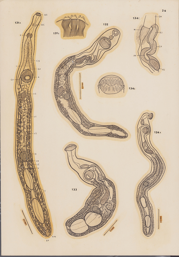
Plate 26
Fig.131 Pseudacaenodera cristata Yamaguti, 1965. A, holotype, ventral view; B, cervical armature, lateral view.
Fig.132 Tormopsolus hawaiiensis n. sp., holotype, dorsal view.
Fig.133 Stephanostomum nunu n. sp., holotype, ventral view.
Fig.134 Stephanostomum hawaiiense n. sp. A, holotype, ventral view; B, circumoral spines; C, terminal genitalia of holotype, ventral view.
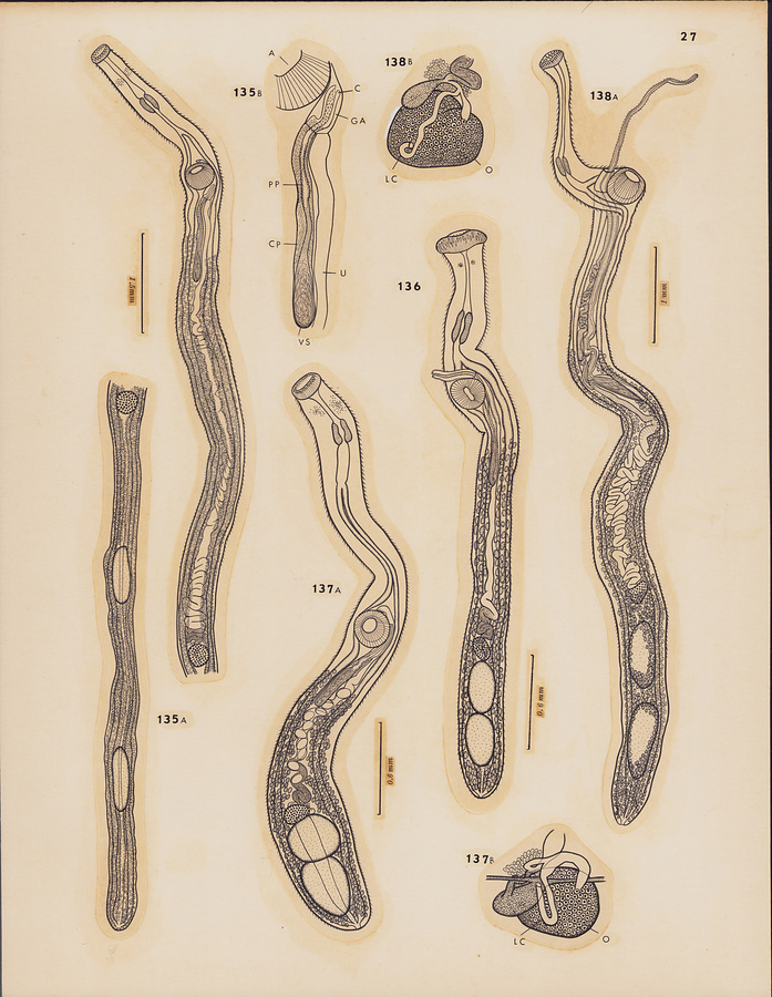
Plate 27
Fig.135 Stephanostomum kawalea n. sp. A, holotype, ventral view; B, terminal genitalia of holotype, ventral view.
Fig.136 Stephanostomum petimba n. sp., holotype, ventral view.
Fig.137 Stephanostomum polymixiae n. sp. A, holotype, ventral view; B, ovarian complex of holotype, dorsal view.
Fig.138 Stephanostomum seriolae n. sp. A, holotype, forebody in ventral view,
hindbody in dorsal view; B, ovarian complex of paratype, dorsal view.
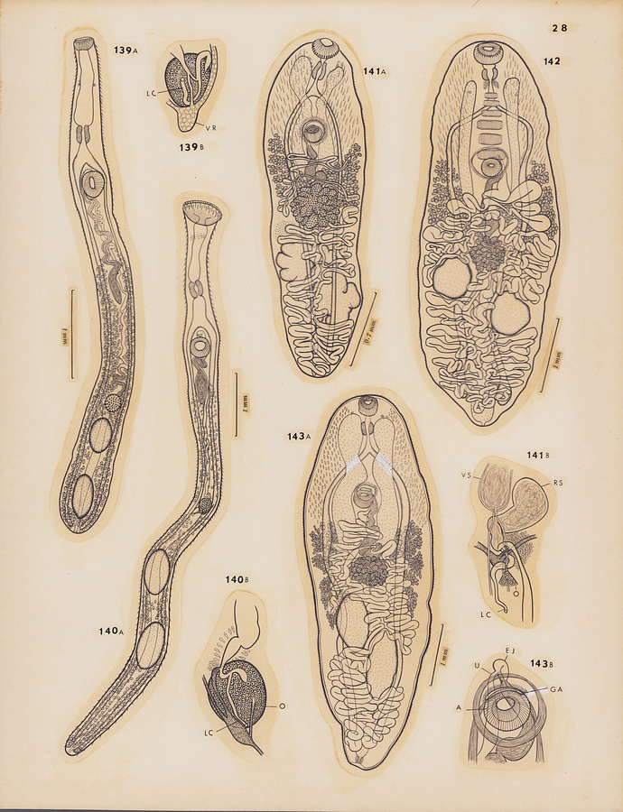
Plate 28
Fig.139 Stephanostomum uku n. sp. A, holotype, ventral view; B, ovarian complex of paratype, dorsal view.
Fig.140 Stephanostomum yagara n. sp. A, holotype, ventral view; B, ovarian complex of holotype, ventral view.
Fig.141 Paracryptogonimus apharei n. sp. A, holotype, ventral view; B, ovarian complex of holotype, ventral view.
Fig.142 Paracryptogonimus muscularis n. sp., holotype, ventral view.
Fig.143 Paracryptogonimus onaga n. sp. A, holotype, ventral view; B, terminal genitalia of paratype, ventral view.
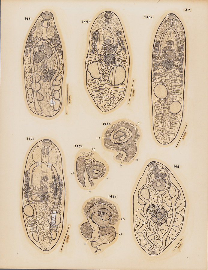
Plate 29
Fig.144 Paracryptogonimus ula-ula n. sp. A, holotype, entral view; B, terminal genitalia of paratype, ventral view.
Fig.145 Siphodera cirrhiti n. sp., holotype, dorsal view.
Fig.146 Pseudosiphoderoides opakapaka n. sp. A, holotype, ventral view; B, terminal genitalia of holotype, ventral view.
Fig.147 Pseudosiphoderoides rooseveltiae n. sp. A, holotype, ventral view; B, terminal genitalia of paratype, ventral view.
Fig.148 Albulatrema ovale Yamaguti, 1965, holotype, ventral view.
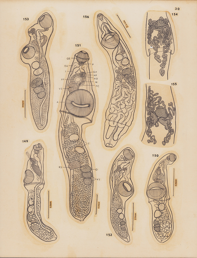
Plate 30
Fig.149 Bunocotyle mugilis n. sp., holotype, lateral view.
Fig.150 Pseudobunocotyla ampladena (Manter et Pritchard, 1960) n. comb., entire worm from Acanthurus dussumieri, ventral view.
Fig.151 Pseudobunocotyla awa Yamaguti, 1965, holotype, ventral view.
Fig.152 Genolinea kyphosi n. sp., holotype, ventral view.
Fig.153 Duosphincter zancli Manter et Pritchard, 1960, entire worm from Zanclus canescens, dorsolateral view.
Fig.154 Dinurus longisinus Looss, 1907, ovariovitellarian region, ventral view.
Fig.155 Lecithocladium chingi Manter et Pritchard, 1960, ovariovitellarian region of a specimen from Naso hexacanthus, ventral view.
Fig.156 Ectenurus lepidus Looss, 1907, entire worm from Decapterus pinnulatus, ventral view.
所蔵館のウェブサイトで見る
公益財団法人 目黒寄生虫館文化庁 〒602-8959 京都府京都市上京区下長者町通新町西入藪之内町85番4
(C) The Agency for Cultural Affairs