ハワイ産魚類の二生吸虫類 原図 9/9
はわいさんぎょるいのにせいきゅうちゅうるい げんず きゅうぶんのきゅう

概要
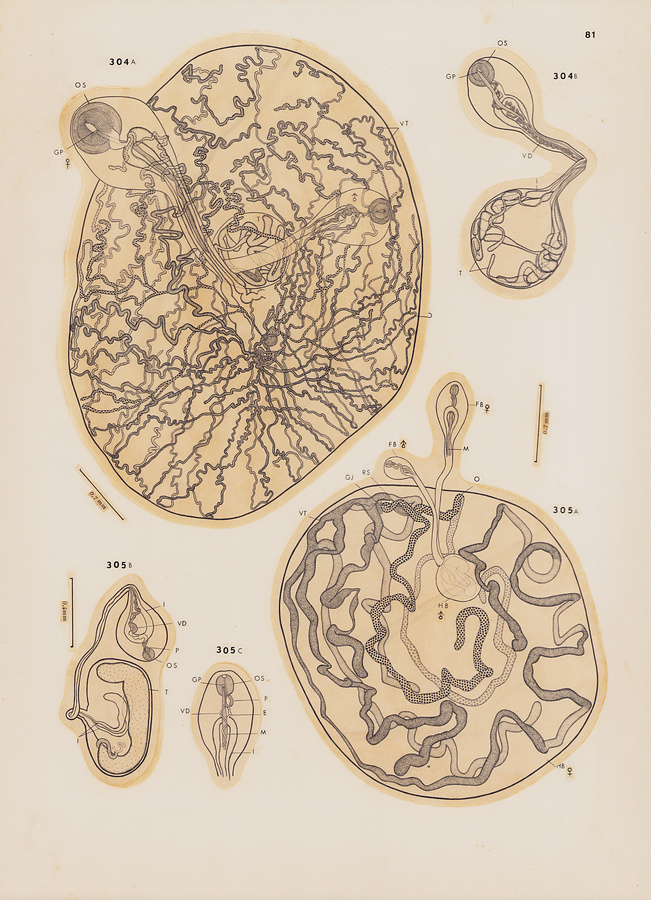
"Plate 81
Fig.304 Koellikeria submaxillaris n. sp. A, holotype, hindbody in ventral view (uterus omitted), forebody of male in ventral view, forebody of female in dorsal view; B, male paratype, ventral view.
Fig.305 Koellikerioides internogastricus n. g., n. sp. A, holotype, hindbody in ventral view, male forebody in ventral view, female forebody in dorsal view; B, male paratype, forebody in dorsal view, hindbody in lateral view; C, anterior extremity of female paratype, ventral view.
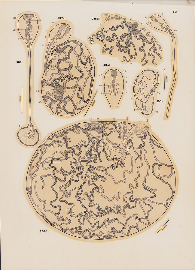
Plate 82
Fig.306 Koellikerioides externogastricus n. sp. A, holotype, ventral view (uterus omitted); B, male paratype, forebody in dorsal view, hindbody in lateral view; C, anterior extremity of female paratype, ventral view; D, region of genital junction of paratype, lateral view (uterus omitted).
Fig.307 Koellikerioides intestinalis n. sp. A, female holotype, ventral view; B, male holotype, forebody in ventral view, hindbody in lateral view.
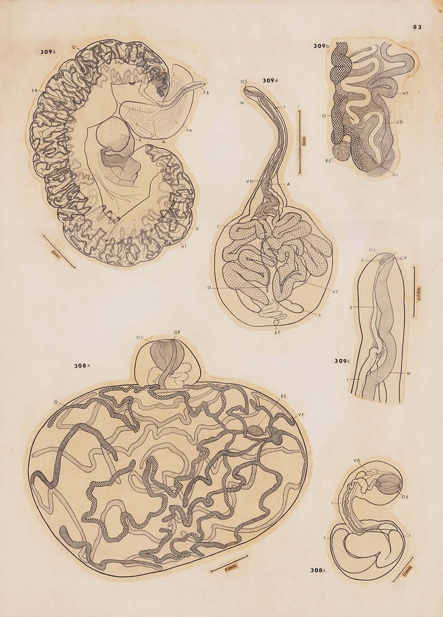
Plate 83
Fig. 308 Koellikerioides apicalis n. sp. A, female holotype, ventral view; B, male holotype, lateral view.
Fig. 309 Patellokoellikeria seriolae n. g., n. sp. A, male holotype, dorsal view; B, female holotype, lateral view; C, anterior extremity of forebody of female paratype, lateral view; D, region of genital junction of female paratype, lateral view.
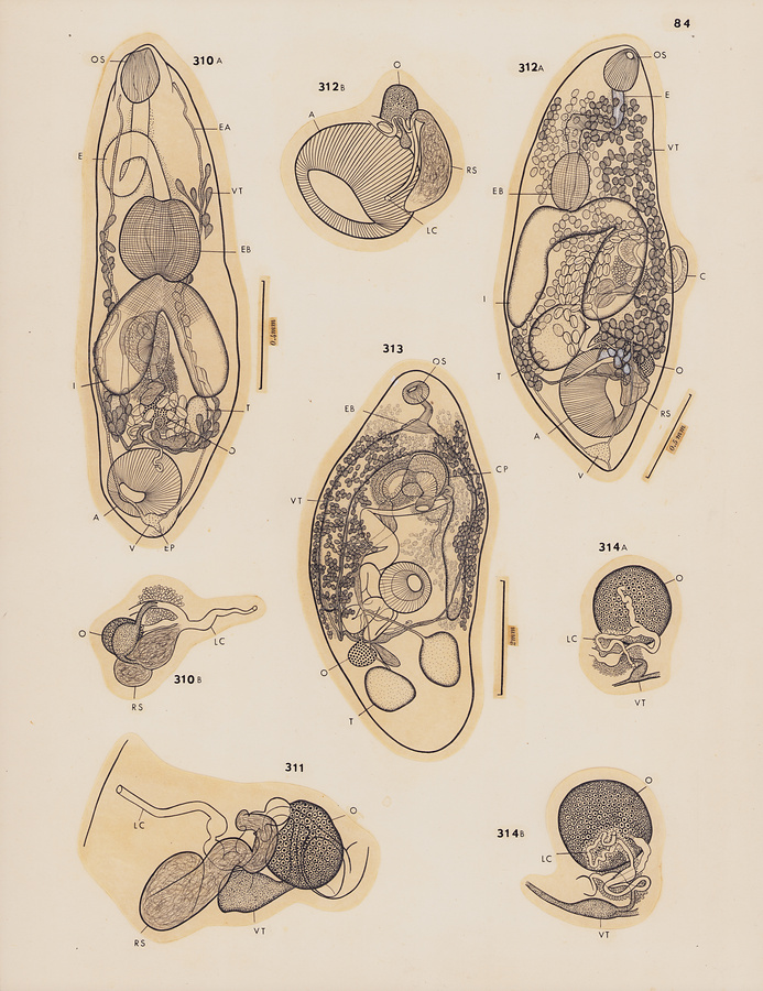
Plate 84
Fig.310 Flagellotrema centropygis n. sp. A, holotype, dorsal view; B, ovarian complex of paratype, lateral view.
Fig.311 Flagellotrema chaetodontis (Manter et Pritchard, 1962) n. comb., ovarian complex of paratype, dorsal view.
Fig.312 Flagellotrema potteri n. sp. A, holotype, dorsal view; B, ovarian complex of paratype, dorsal view.
Fig.313 Leptobulbus magnacirratus Manter et Pritchard, 1962, from Scarus sordidus, ventral view.
Fig.314 Cleptodiscus bulbosus Hanson, 1955. A & B, ovarian complexes showing different courses of Laurer's canal, probably due to different degrees of pressure applied on cover glass.
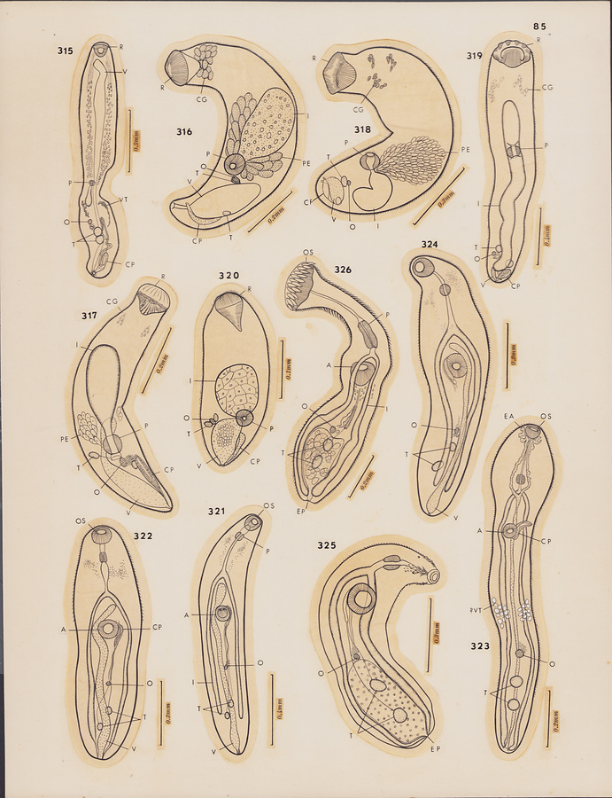
Plate 85
Fig.315 Larval form of Rhipidocotyle sp. from Pranesus insularum, ventral view.
Fig.316 Larval form of Rhipidocotyle capitata (Linton, 1940) from Auxis thazard, ventral view.
Fig.317 Larval form of Dollfustrema sp. from fin-ray of Amanses pardalis, ventral view.
Fig.318 Larval form of Rhipidocotyle kawakawa n. sp. from gill of Cypselurus spilonotopterus, lateral view.
Fig.319 Larval form of Dolichoenterum congeri n. sp. from visceral wall of Istioblennius zebra, ventral view.
Fig.320 Larval form of Prosorhynchus sp. from fin-ray of Mulloidichthys auriflamma, ventral view.
Fig.321 Encysted metacercaria of Lepidapedon sp. free in stomach of Odontanthias fuscipinnis, ventral view.
Fig.322 Larval form of Preptetos sp. or Lepidapedon sp. from intestine of Chromis verater, ventral view.
Fig.323 Larval form of Allolepidapedon petimba n. sp. from dorsal muscle of Thalassoma umbrostigma, ventral view.
Fig.324 Larval form of lepocreadiid from Merinthe macrocephala, dorsal view.
Fig.325 Larval form of Acaenodera placophora Manter et Pritchard, 1960, from muscle of Thalassoma umbrostigma, ventral view.
Fig.326 Larval form of Stephanostomum sp. with 31-32 circumoral spines, from Alutera scripta, dorsal view.
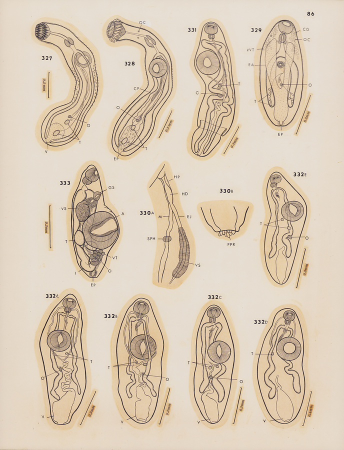
Plate 86
Fig.327 Larval form of Stephanostomum sp. with 35-37 circumoral spines, from Diodon holocanthus, dorsal view.
Fig.328 Larval form of Stephanostomum sp. with 36 circumoral spines, from Zanclus canescens, ventral view.
Fig.329 Larval form of Siphodera cirrhiti n. sp. from Istioblennius zebra, ventral view.
Fig.330 Larval form of Musculovesicula bilabiata Manter et Pritchard, 1960 from stomach wall of Abudefduf sp. A, terminal genitalia; B, posterior extremity, lateral view.
Fig.331 Larval form of Ectenurus sp. from Elagatis bipinnulatus, ventral view.
Fig.332 Unidentifiable encysted hemiurid larval forms. A, from stomach wall of Synodus dermatogenys, ventral view; B, from subserosa of gastrointestinal tract of Anampses godeffroyi, dorsal view; C, from mesentery of Gomphosus varius, ventral view; D, from body cavity and muscle of Thalassoma umbrostigma, ventral view; E, from muscle of Bathygobius fuscus, ventral view.
Fig.333 Juvenile form of Lobatovitelliovarium fusiforme Yamaguti, 1965, from muscle of Pranesus insularum, ventrolateral view.
Plate 87
Fig.334 Monilicaecum from Bathygobius fuscus, dorsal view.
Fig.335 Torticaecum of Allonematobothrium apharei n. sp. from Aphareus rutilans, ventral view.
Fig.336 Posttorticaecum (A) and juvenile (B) of Didymocystis acanthocybii Yamaguti, 1938, from Acanthocybium solandri.
Fig.337 Postmonilicaecum from subserosa of stomach of Auxis thazard, ventral view.
Fig.338 Later Posttorticaecum stage of Metadidymocystis cymbiformis found in washings of stomach wall of Lampris regius, ventral view.
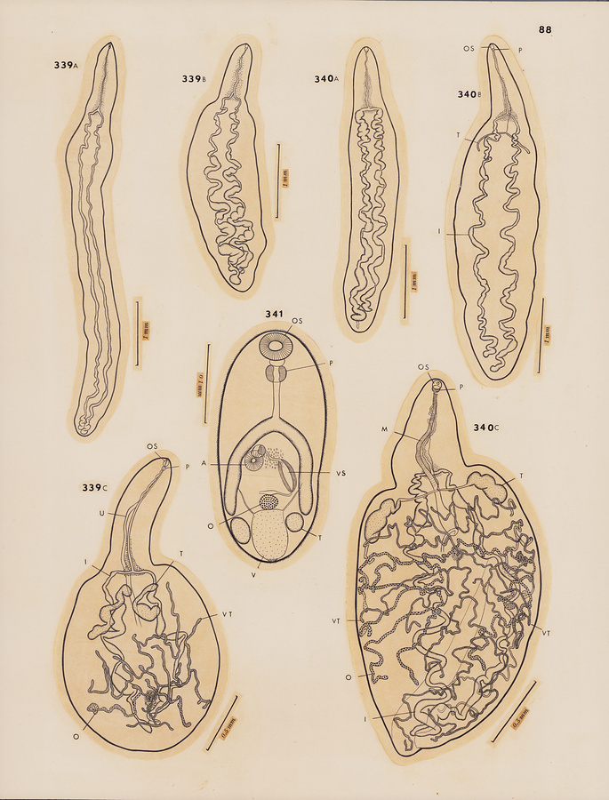
Plate 87
Fig.339 Larval stages of Didymocystis irregularis n. sp. from Neothunnus macropterus. A, Posttorticaecum, ventral view; B, intermediate stage between Posttorticaecum and juvenile form, ventral view; C, juvenile form, dorsal view.
Fig.340 Larval stages of Didymocystis superpalati n. sp. from Neothunnus macropterus. A, Postmonilicaecum, ventral view; B, intermediate stage between Postmonilicaecum and juvenile form, dorsal view; C, juvenile form, dorsal view.
Fig.341 Metacercaria of Stellantchasmus falcatus Onji et Nishio, 1915, from muscle of Mugil cephalus, ventral view.
所蔵館のウェブサイトで見る
公益財団法人 目黒寄生虫館文化庁 〒602-8959 京都府京都市上京区下長者町通新町西入藪之内町85番4
(C) The Agency for Cultural Affairs