ハワイ産魚類の二生吸虫類 原図 5/9
はわいさんぎょるいのにせいきゅうちゅうるい げんず きゅうぶんのご
概要
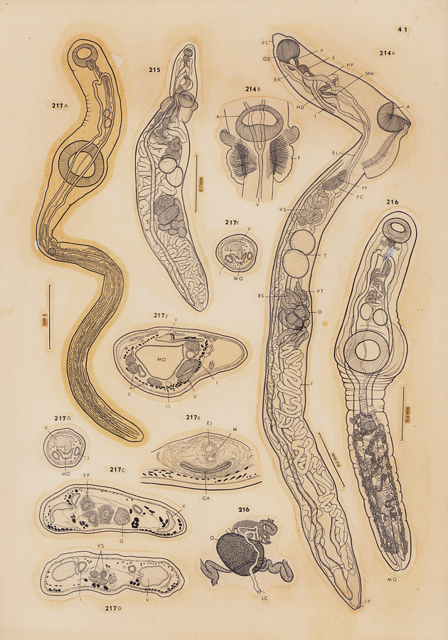
Plate 41
Fig.214 Quadrifoliovarium pritchardae Yamaguti, 1965. A, holotype, lateral view; B, two separate postacetabular flaps of paratype, ventral view.
Fig.215 Holacanthitrema lobatum n. g., n. sp., holotype, ventral view.
Fig.216 Prosorchiopsis aluterae n. sp. A, holotype, ventral view; B, ovarian complex of holotype, dorsal view.
Fig.217 Prosorchiopsis nasonis n. sp. A, holotype, dorsal view; B-G, transverse sections through genital atrium, pars prostatica, vesicula seminalis, and Manter's organ, respectively.
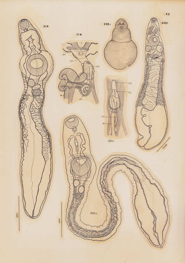
Plate 42
Fig.218 Sclerodistomum bravoae Pritchard, 1963, terminal genitalia of a specimen from Diodon sp., ventral view.
Fig.219 Hirudinella ahi n. sp., holotype from Neothunnus macropterus, ventral view.
Fig.220 Hirudinella beebei Chandler, 1937. A, entire worm (contracted), ventral view; B, same (extended), ventral view.
Fig.221 Hirudinella marina (Garcin, 1730). A, entire worm from Katsuwonus pelamys, ventral view; B, terminal genitalia, ventral view.
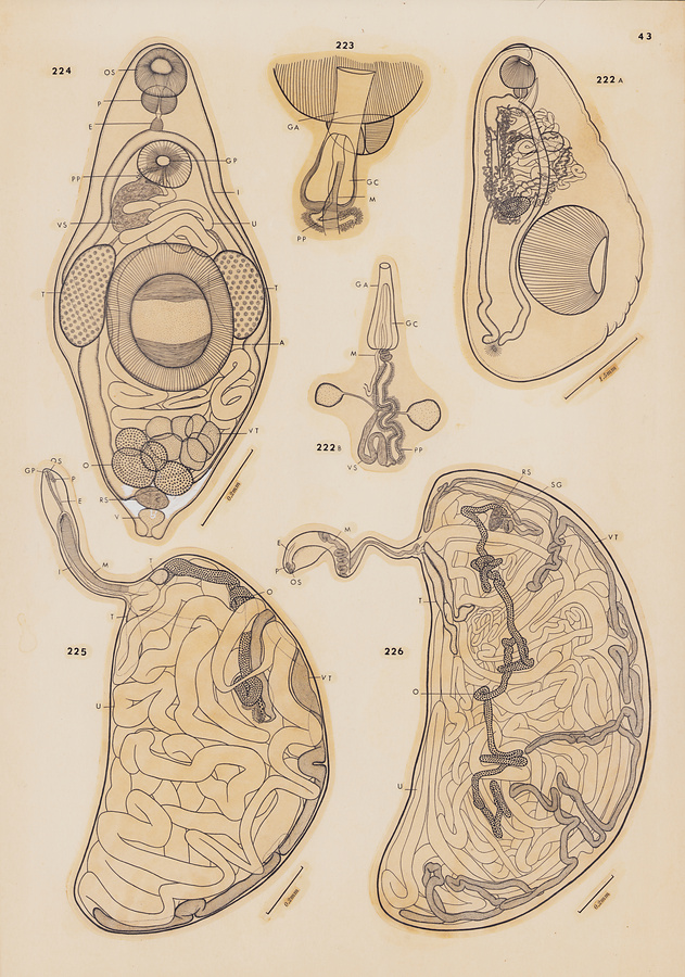
Plate 43
Fig.222 Prosogonotrema symmetricum Oshmarin, 1965. A, specimen from Pristipomoides microlepis; dorsolateral view; B, male genitalia, ventral view.
Fig.223 Prosogonotrema subequilatum Pritchard, 1963, terminal genitalia of a specimen from Naso hexacanthus, ventral view.
Fig.224 Lobatovitelliovarium fusiforme Yamaguti, 1965, holotype, ventral view.
Fig.225 Dermatodidymocystis vivipara n. g., n. sp., holotype, from Parathunnus sibi; forebody in ventral view, hindbody in lateral view.
Fig.226 Dermatodidymocystis viviparoides n. sp., holotype from Neothunnus macropterus, lateral view.
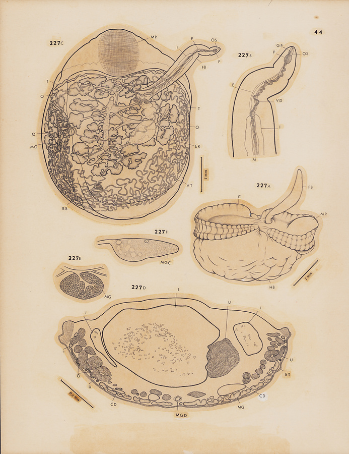
Plate 44
Fig.227 Adenodidymocystis intestinalis n. g., n. sp. A, entire paratype, ventrolateral view; B, anterior extremity of forebody of holotype, ventral view; C, holotype,ventral view; D, transverse section through common duct of mucoid glands; E, mucoid gland; F, a large mucoid gland cell, apparently active in function.
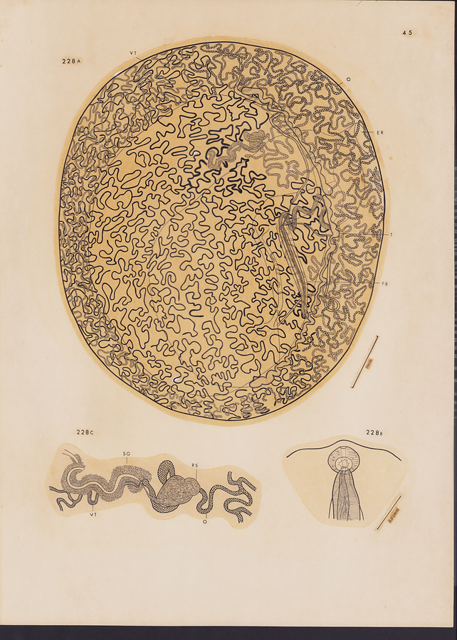
Plate 45
Fig.228 Coeliodidymocystis kamegaii n. g., n. sp. A, holotype, ventral view (uterus omitted except for metraterm); B, head end of paratype, ventral view; C, ovarian complex of holotype, ventral view.
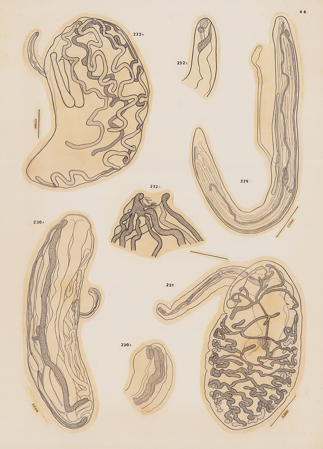
Plate 46
Fig.229 Didymocylindrus filiformis Ishii, 1935, entire worm from Katsuwonus pelamys, lateral view.
Fig.230 Didymocylindrus simplex (Ishii, 1935) n. comb. A, entire worm from Katsuwonus pelamys, lateral view; B, anterior extremity of same, lateral view.
Fig.231 Didymocystis acanthocybii Yamaguti, 1938, entire worm, dorsal view.
Fig.232 Didymocystis bifurcata n. sp. A, holotype, lateral view; B, anterior extremity of paratype, lateral view; C, genital junction of paratype, lateral view.
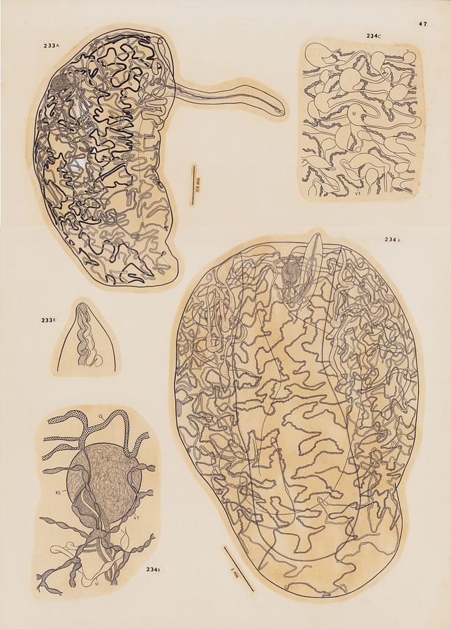
Plate 47
Fig.233 Didymocystis irregularis n. sp. A, holotype from Neothunnus macropterus, lateral view; B, anterior extremity of paratype, ventral view.
Fig.234 Didymocystis nasalis n. sp. A, holotype, ventral view; B, ovarian complex of holotype; C, moniliform uterus and moniliform vitelline gland in holotype, ventral view.
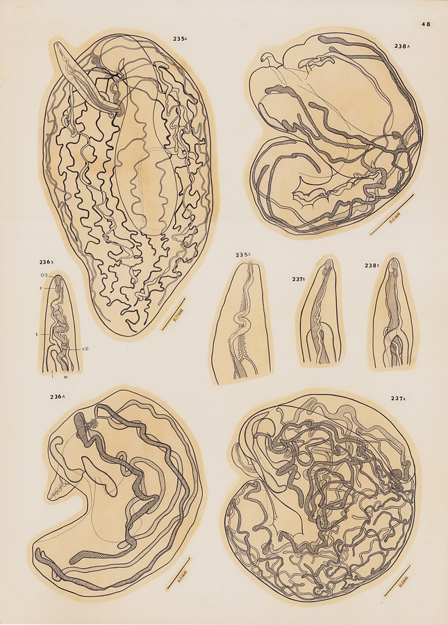
Plate 48
Fig.235 Didymocystis orbitalis n. sp. A, holotype, ventral view; B, anterior extremity of holotype, ventral view.
Fig.236 Didymocystis palati n. sp. A, holotype, lateral view; B, anterior extremity of paratype, ventral view.
Fig.237 Didymocystis philobranchia n. sp. A, holotype from Neothunnus macropterus, lateral view; B, anterior extremity of paratype, ventral view.
Fig.238 Didymocystis philobranchiarca n. sp. A, holotype, lateral view; B, anterior extremity of paratype, dorsal view.
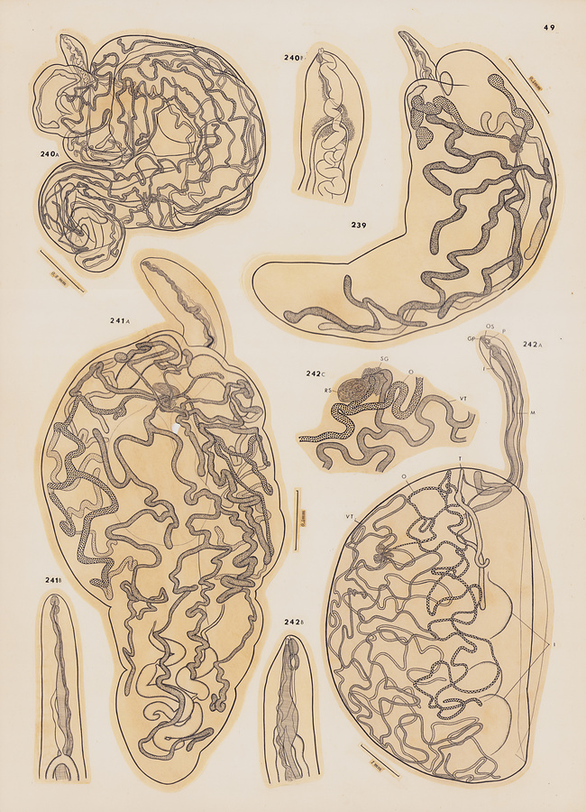
Plate 49
Fig.239 Didymocystis poonui n. sp., holotype, dorsolateral view.
Fig.240 Didymocystis spirocauda n. sp. A, holotype, forebody in ventral view, hindbody in lateral view; B, anterior extremity of holotype, ventral view.
Fig.241 Didymocystis superpalati n. sp. A, holotype, dorsal view; B, anterior extremity of paratype, ventral view.
Fig.242 Didymocystoides bifasciatus n. g., n. sp. A, holotype, forebody in ventral view, hindbody in lateral view; B, anterior extremity of paratype, ventral view; C, ovarian complex of paratype, lateral view.
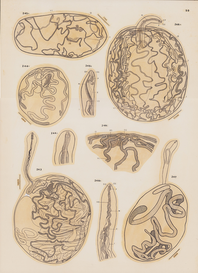
Plate 50
Fig.243 Didymocystoides buccalis n. sp., holotype, forebody in ventral view, hindbody in ventrolateral view.
Fig.244 Didymocystoides exiguus n. sp. A, holotype, dorsal view; B, anterior extremity of paratype, ventral view.
Fig.245 Didymocystoides intestinomuscularis n. sp. A, holotype, dorsal view; B, anterior extremity of paratype, ventrolateral view.
Fig.246 Didymocystoides oesophagicola n. sp. A, holotype, ventral view; B, anterior extremity of paratype, ventral view; C, ovarian complex of paratype, dorsal view.
Fig.247 Didymocystoides pectoralis n. sp., holotype, dorsal view.
所蔵館のウェブサイトで見る
公益財団法人 目黒寄生虫館文化庁 〒602-8959 京都府京都市上京区下長者町通新町西入藪之内町85番4
(C) The Agency for Cultural Affairs