ハワイ産魚類の二生吸虫類 原図 1/9
はわいさんぎょるいのにせいきゅうちゅうるい げんず きゅうぶんのいち
概要
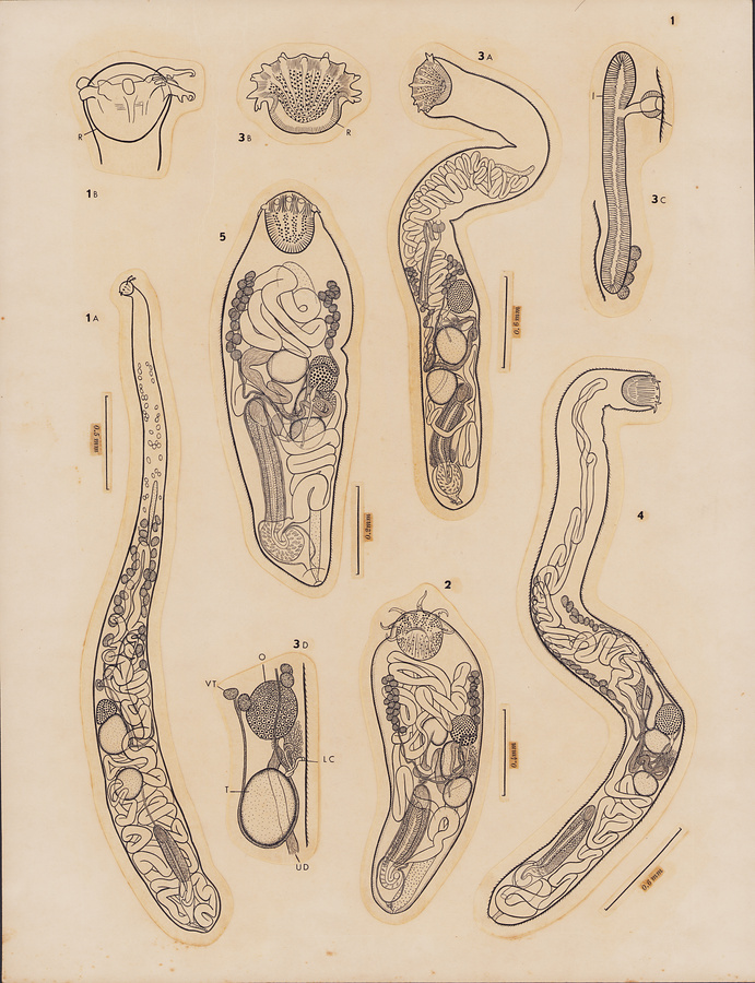
Plate 1
Fig.1 Bucephalus carangis n. sp. A, holotype, ventral view; B, anterior extremity of holotype, ventral view.
Fig.2 Bucephalus carangoides n. sp., holotype,dorsal view.
Fig.3 Bucephalus kaku n. sp. A, holotype, dorsal view; B, rhynchus of paratype from type host, lateral view; C, pharynx, esophagus and intestine of paratype from type host, lateral view; D, ovarian complex of paratype from Sphyraena helleri, lateral view.
Fig.4 Bucephalus sextentaculatus n. sp., holotype, dorsal view.
Fig.5 Bucephalus ulua n. sp., holotype, dorsal view.
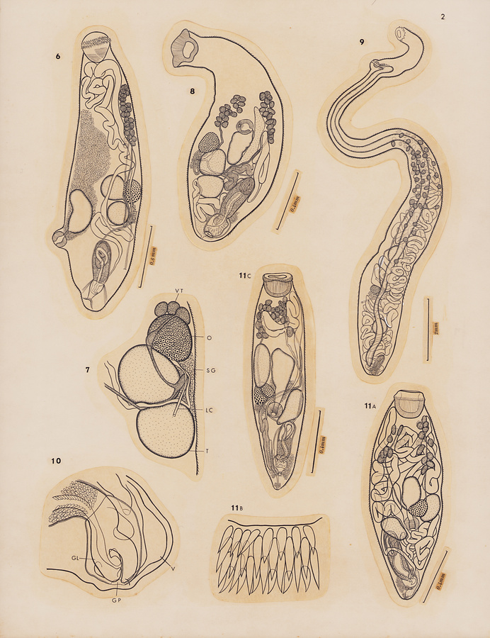
Plate 2
Fig.6 Dollfustrema stromborhynchum Manter et Pritchard, 1961, lateral view.
Fig.7 Rhipidocotyle capitata (Linton, 1940), testes and ovarian complex, dorsal view.
Fig.8 Rhipidocotyle kawakawa n. sp. holotype, ventral view.
Fig.9 Dolichoenterum congeri n. sp., holotype, ventrolateral view.
Fig.10 Bucephalopsis arcuata (Linton, 1900), posterior extremity, dorsal view.
Fig.11 Bucephalopsis bipapillosa (Manter et Pritchard, 1961) n. comb.A, entire worm from Gymnothorax undulatus, dorsal view; B, part of rhynchus of a specimen from Gymnothorax undulatus, ventral view; C, a specimen from Gymnothorax petelli (type host), ventral view.
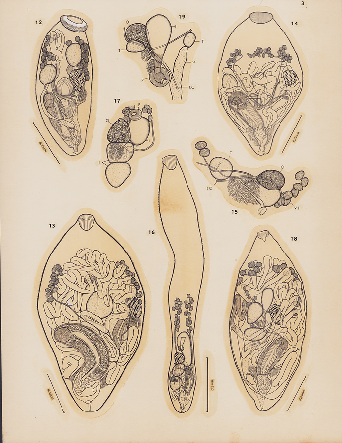
Plate 3
Fig.12 Bucephalopsis puhilaumilo n. sp., holotype, dorsal view.
Fig.13 Prosorhynchus berycis n. sp., holotype, dorsal view.
Fig.14 Prosorhynchus congeri n. sp., holotype, dorsal view.
Fig.15 Prosorhynchus epinepheli Yamaguti, 1939, ovarian complex and vicinity, dorsal view.
Fig.16 Prosorhynchus kahala n. sp., holotype, ventral view.
Fig.17 Prosorhynchus longicollis Yamaguti, 1953, ovarian complex, ventral view.
Fig.18 Prosorhynchus polydactyli n. sp., holotype, ventral view.
Fig.19 Prosorhynchus uniporus Ozaki, 1924, ovarian complex and vicinity, ventral view.
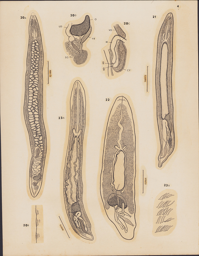
Plate 4
Fig.20 Aporocotyle pacifica n. sp. A, holotype, dorsal view; B, longitudinal groups of spines on ventrolateral margin of body; C, terminal genitalia of holotype, dorsal view; D, ovarian complex, dorsal view.
Fig.21 Cardicola ahi n. sp., holotype from Neothunnus macropterus, dorsal view.
Fig.22 Cardicola chaetodontis n. sp., holotype from Chaetodon miliaris, dorsal view.
Fig.23 Cardicola mugilis n. sp. A, holotype, ventral view; B, longitudinal series of transverse rows of spines on ventral submargin of body.
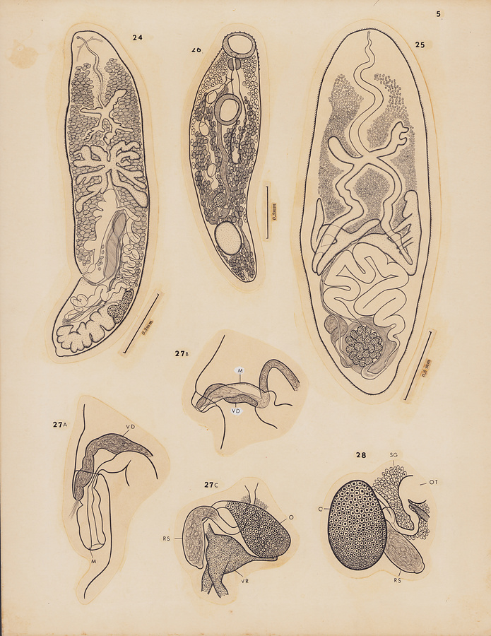
Plate 5
Fig.24 Neoparacardicola nasonis n. g., n. sp., holotype, dorsal view.
Fig.25 Deontacylix kyphosi n. sp., holotype, ventral view.
Fig.26 Schikhobalotrema acanthuri n. sp., holotype, ventral view.
Fig.27 Schikhobalotrema hawaiiense Pritchard et Manter, 1961. A, terminal genitalia, completely evaginated, lateral view; B, same, partly protruded, lateral view; C, ovarian complex, lateral view.
Fig.28
Schikhobalotrema robustum Pritchard et Manter, 1961, ovarian complex, ventral view.
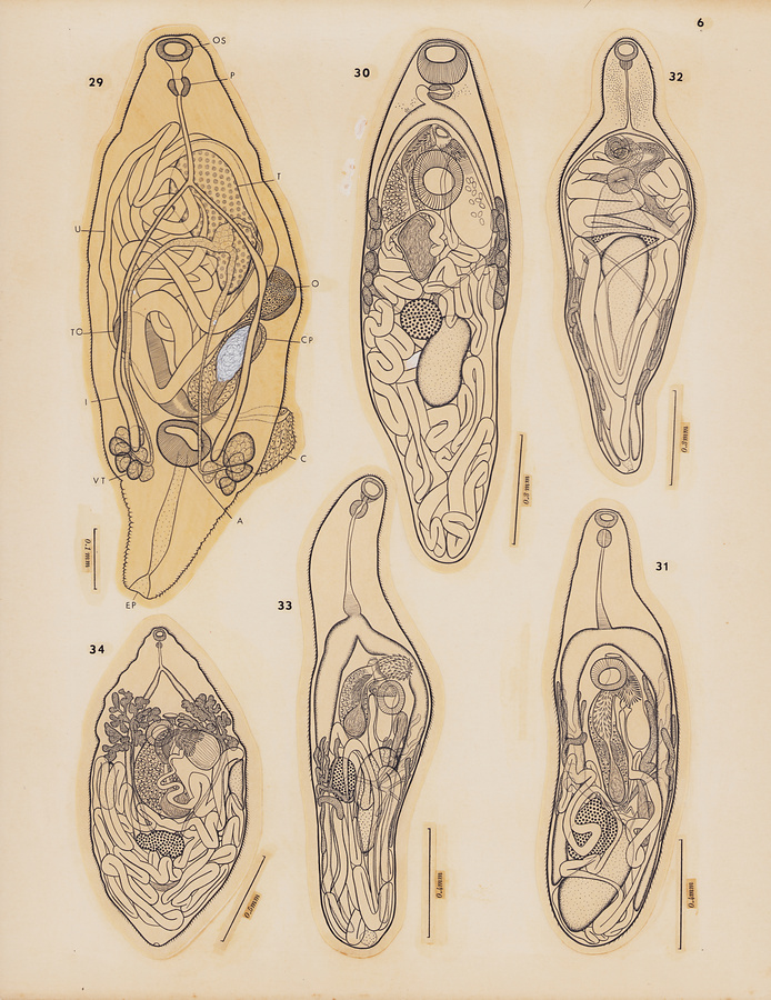
Plate 6
Fig.29 Anamonorchis ulua n. g., n. sp., holotype, dorsal view.
Fig.30 Lasiotocus oculatus (Manter et Pritchard, 1961), ventral view.
Fig.31 Lasiotocus delicatus Manter et Pritchard, 1961, from Parupeneus multifasciatus, ventral view.
Fig.32 Lasiotocus ulua n. sp., holotype, ventral view.
Fig.33 Lasiotocus weke n. sp., holotype from Mulloidichthys samoensis, ventral view.
Fig.34 Paramonorcheides spicularis n. sp., holotype from Decapterus pinnulatus, ventral view.
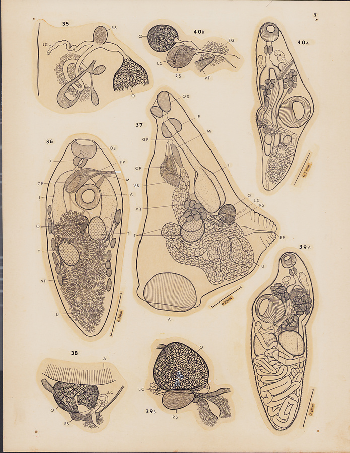
Plate 7
Fig.35 Hurleytrematoides coronatus Manter et Pritchard, 1961, from Chaetodon fremblii, ovarian complex, lateral view.
Fig.36 Cypseluritrema spilonotopteri n. g., n. sp., holotype, ventral view.
Fig.37 Cypseluritrematoides triangularis n. g., n. sp., holotype, lateral view.
Fig.38 Deretrema acutum Pritchard, 1963, from Naso hexacanthus, Ovarian complex, ventral view.
Fig.39 Deretrema carangis n. sp. A, holotype, ventral view; B, ovarian complex, dorsal view.
Fig.40 Deretrema hawaiiense n. sp. A, holotype from Elagatis bipinnulatus, ventral view; B, ovarian complex of holotype, ventral view.
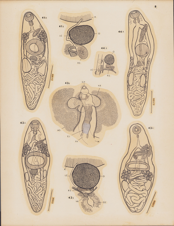
Plate 8
Fig.41 Deretrema sphyraenae n. sp. A, holotype, ventral view; B, ovarian complex, dorsal view.
Fig.42 Deretrema uku n. sp. A, holotype from Aprion virescens, ventral view; B, ovarian complex, dorsal view.
Fig.43 Proctophantastes polymixiae n. sp. A, holotype, ventral view; B, terminal genitalia of paratype, ventral view.
Fig.44 Lecithostaphylus aha-aha n. sp. A, holotype, ventral view; B, ovarian complex of paratype, dorsal view.
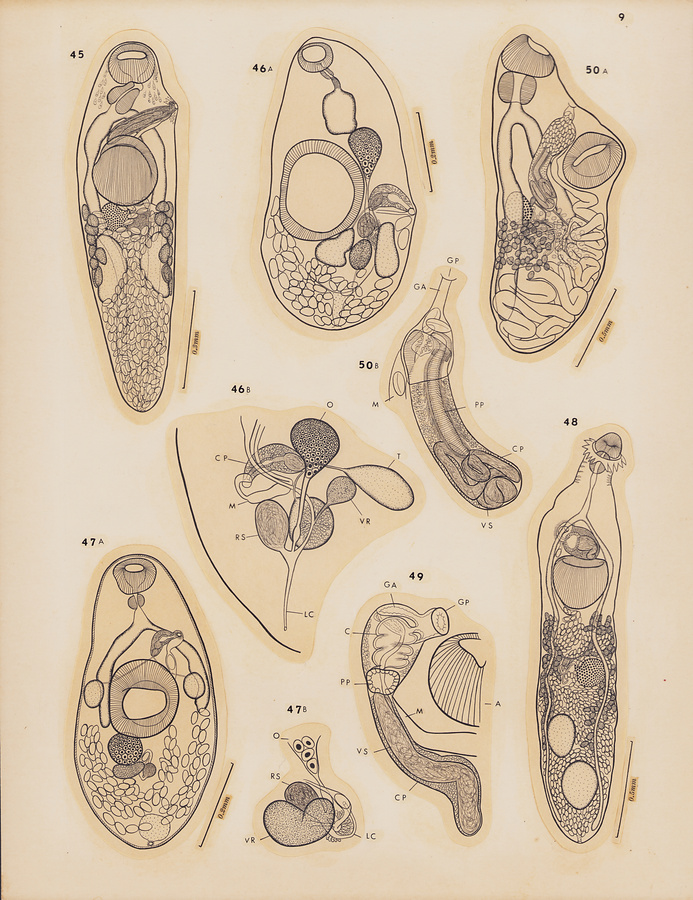
Plate 9
Fig.45 Lecithostaphylus depauperati n. sp., holotype, ventral view.
Fig.46 Parvipyrum acanthuri Pritchard, 1963. A, entire worm from Acanthurus olivaceus, ventral view; B, ovarian complex and terminal genitalia of a specimen from Acanthurus dussumieri, lateral view.
Fig.47 Zoogonoides synodi n. sp. A, holotype, ventral view; B, ovarian complex of paratype, ventral view.
Fig.48 Tergestia kuhliae n. sp., holotype, dorsal view.
Fig.49 Tergestia laticollis (Rud., 1819) Odhner, 1911, terminal genitalia of a specimen from Decapterus pinnulatus, ventral view.
Fig.50 Proctoeces hawaiiensis n. sp. A, holotype, ventrolateral view; B, terminal genitalia of paratype, ventral view.
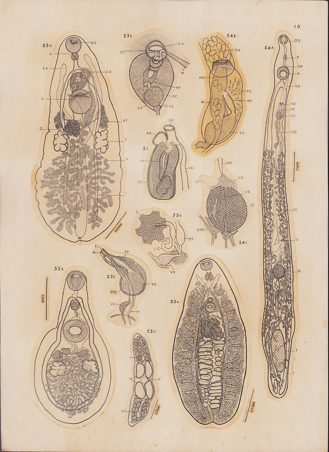
Plate 10
Fig.51 Lintonium consors (Lühe, 1906) Crowcroft, 1950, terminal genitalia ventral view.
Fig.52 Xystretrum hawaiiense n. sp. A, holotype from Balistes capistratus ventral view; B, terminal genitalia of paratype, ventral view.
Fig.53 Callodistomoides foliatus n. g., n. sp. A, holotype, ventral view; terminal genitalia, ventral view.
Fig.54 Hapladena nasonis n. sp. A, holotype, ventral view; B, terminal genital of paratype, ventral view; C, ovarian complex of holotype, ventral view.
Fig.55 Pleorchis uku n. sp. A, holotype, ventral view; B, ovarian complex of paratype, ventral view; C, transverse section of paratype showing arrangement of testes.
所蔵館のウェブサイトで見る
公益財団法人 目黒寄生虫館文化庁 〒602-8959 京都府京都市上京区下長者町通新町西入藪之内町85番4
(C) The Agency for Cultural Affairs